Iron »
PDB 3whm-3x16 »
3wkt »
Iron in PDB 3wkt: Complex Structure of An Open Form of Nadph-Cytochrome P450 Reductase and Heme Oxygenase-1
Enzymatic activity of Complex Structure of An Open Form of Nadph-Cytochrome P450 Reductase and Heme Oxygenase-1
All present enzymatic activity of Complex Structure of An Open Form of Nadph-Cytochrome P450 Reductase and Heme Oxygenase-1:
1.14.99.3; 1.6.2.4;
1.14.99.3; 1.6.2.4;
Protein crystallography data
The structure of Complex Structure of An Open Form of Nadph-Cytochrome P450 Reductase and Heme Oxygenase-1, PDB code: 3wkt
was solved by
M.Sugishima,
H.Sato,
Y.Higashimoto,
J.Harada,
K.Wada,
K.Fukuyama,
M.Noguchi,
with X-Ray Crystallography technique. A brief refinement statistics is given in the table below:
| Resolution Low / High (Å) | 41.34 / 4.30 |
| Space group | P 61 |
| Cell size a, b, c (Å), α, β, γ (°) | 290.336, 290.336, 83.646, 90.00, 90.00, 120.00 |
| R / Rfree (%) | 22.2 / 25.6 |
Iron Binding Sites:
The binding sites of Iron atom in the Complex Structure of An Open Form of Nadph-Cytochrome P450 Reductase and Heme Oxygenase-1
(pdb code 3wkt). This binding sites where shown within
5.0 Angstroms radius around Iron atom.
In total 2 binding sites of Iron where determined in the Complex Structure of An Open Form of Nadph-Cytochrome P450 Reductase and Heme Oxygenase-1, PDB code: 3wkt:
Jump to Iron binding site number: 1; 2;
In total 2 binding sites of Iron where determined in the Complex Structure of An Open Form of Nadph-Cytochrome P450 Reductase and Heme Oxygenase-1, PDB code: 3wkt:
Jump to Iron binding site number: 1; 2;
Iron binding site 1 out of 2 in 3wkt
Go back to
Iron binding site 1 out
of 2 in the Complex Structure of An Open Form of Nadph-Cytochrome P450 Reductase and Heme Oxygenase-1
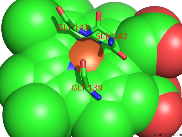
Mono view
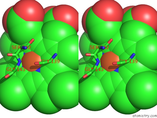
Stereo pair view
Mono view
Stereo pair view
A full contact list of Iron with other atoms in the Fe binding
site number 1 of Complex Structure of An Open Form of Nadph-Cytochrome P450 Reductase and Heme Oxygenase-1 within 5.0Å range:
|
Iron binding site 2 out of 2 in 3wkt
Go back to
Iron binding site 2 out
of 2 in the Complex Structure of An Open Form of Nadph-Cytochrome P450 Reductase and Heme Oxygenase-1
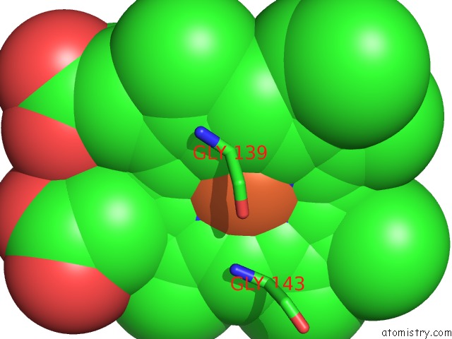
Mono view
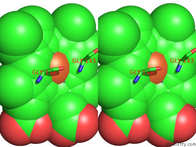
Stereo pair view
Mono view
Stereo pair view
A full contact list of Iron with other atoms in the Fe binding
site number 2 of Complex Structure of An Open Form of Nadph-Cytochrome P450 Reductase and Heme Oxygenase-1 within 5.0Å range:
|
Reference:
M.Sugishima,
H.Sato,
Y.Higashimoto,
J.Harada,
K.Wada,
K.Fukuyama,
M.Noguchi.
Structural Basis For the Electron Transfer From An Open Form of Nadph-Cytochrome P450 Oxidoreductase to Heme Oxygenase To Be Published.
Page generated: Tue Aug 5 08:23:55 2025
Last articles
Mn in 9LJUMn in 9LJW
Mn in 9LJS
Mn in 9LJR
Mn in 9LJT
Mn in 9LJV
Mg in 9UA2
Mg in 9R96
Mg in 9VM1
Mg in 9P01