Iron »
PDB 4b31-4bmp »
4bmp »
Iron in PDB 4bmp: Crystal Structure of Bacillus Cereus Ribonucleotide Reductase Di-Iron Nrdf in Complex with Nrdi (2.1 A Resolution)
Enzymatic activity of Crystal Structure of Bacillus Cereus Ribonucleotide Reductase Di-Iron Nrdf in Complex with Nrdi (2.1 A Resolution)
All present enzymatic activity of Crystal Structure of Bacillus Cereus Ribonucleotide Reductase Di-Iron Nrdf in Complex with Nrdi (2.1 A Resolution):
1.17.4.1;
1.17.4.1;
Protein crystallography data
The structure of Crystal Structure of Bacillus Cereus Ribonucleotide Reductase Di-Iron Nrdf in Complex with Nrdi (2.1 A Resolution), PDB code: 4bmp
was solved by
M.Hammerstad,
H.-P.Hersleth,
A.K.Rohr,
K.K.Andersson,
with X-Ray Crystallography technique. A brief refinement statistics is given in the table below:
| Resolution Low / High (Å) | 62.59 / 2.10 |
| Space group | C 2 2 21 |
| Cell size a, b, c (Å), α, β, γ (°) | 59.330, 125.030, 142.561, 90.00, 90.00, 90.00 |
| R / Rfree (%) | 17.695 / 23.342 |
Other elements in 4bmp:
The structure of Crystal Structure of Bacillus Cereus Ribonucleotide Reductase Di-Iron Nrdf in Complex with Nrdi (2.1 A Resolution) also contains other interesting chemical elements:
| Chlorine | (Cl) | 1 atom |
Iron Binding Sites:
The binding sites of Iron atom in the Crystal Structure of Bacillus Cereus Ribonucleotide Reductase Di-Iron Nrdf in Complex with Nrdi (2.1 A Resolution)
(pdb code 4bmp). This binding sites where shown within
5.0 Angstroms radius around Iron atom.
In total 2 binding sites of Iron where determined in the Crystal Structure of Bacillus Cereus Ribonucleotide Reductase Di-Iron Nrdf in Complex with Nrdi (2.1 A Resolution), PDB code: 4bmp:
Jump to Iron binding site number: 1; 2;
In total 2 binding sites of Iron where determined in the Crystal Structure of Bacillus Cereus Ribonucleotide Reductase Di-Iron Nrdf in Complex with Nrdi (2.1 A Resolution), PDB code: 4bmp:
Jump to Iron binding site number: 1; 2;
Iron binding site 1 out of 2 in 4bmp
Go back to
Iron binding site 1 out
of 2 in the Crystal Structure of Bacillus Cereus Ribonucleotide Reductase Di-Iron Nrdf in Complex with Nrdi (2.1 A Resolution)
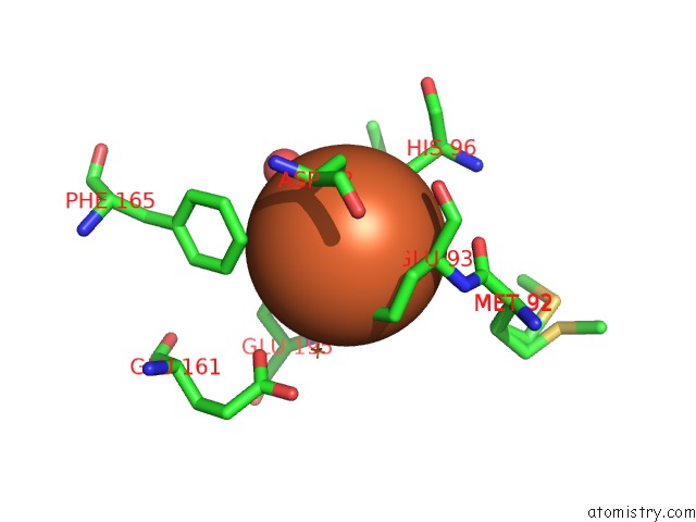
Mono view
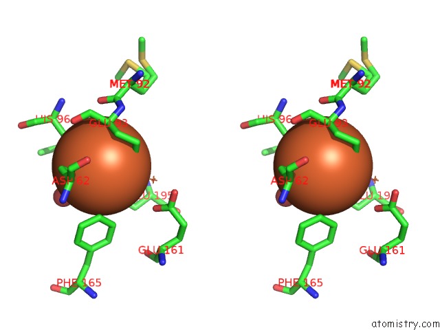
Stereo pair view
Mono view
Stereo pair view
A full contact list of Iron with other atoms in the Fe binding
site number 1 of Crystal Structure of Bacillus Cereus Ribonucleotide Reductase Di-Iron Nrdf in Complex with Nrdi (2.1 A Resolution) within 5.0Å range:
|
Iron binding site 2 out of 2 in 4bmp
Go back to
Iron binding site 2 out
of 2 in the Crystal Structure of Bacillus Cereus Ribonucleotide Reductase Di-Iron Nrdf in Complex with Nrdi (2.1 A Resolution)
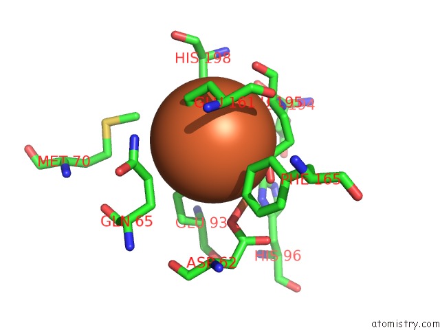
Mono view
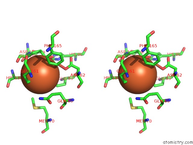
Stereo pair view
Mono view
Stereo pair view
A full contact list of Iron with other atoms in the Fe binding
site number 2 of Crystal Structure of Bacillus Cereus Ribonucleotide Reductase Di-Iron Nrdf in Complex with Nrdi (2.1 A Resolution) within 5.0Å range:
|
Reference:
M.Hammerstad,
H.Hersleth,
A.B.Tomter,
A.K.Rohr,
K.K.Andersson.
Crystal Structure of Bacillus Cereus Class Ib Ribonucleotide Reductase Di-Iron Nrdf in Complex with Nrdi. Acs Chem.Biol. V. 9 526 2014.
ISSN: ISSN 1554-8929
PubMed: 24295378
DOI: 10.1021/CB400757H
Page generated: Tue Aug 5 09:16:58 2025
ISSN: ISSN 1554-8929
PubMed: 24295378
DOI: 10.1021/CB400757H
Last articles
Mg in 1K9WMg in 1K9Y
Mg in 1KAY
Mg in 1KAO
Mg in 1KAX
Mg in 1KA2
Mg in 1KA1
Mg in 1K9M
Mg in 1K8A
Mg in 1K73