Iron »
PDB 4dig-4egm »
4dxu »
Iron in PDB 4dxu: Crystal Structure of C-Lobe of Bovine Lactoferrin Complexed with Aminocaproic Acid at 1.46 A Resolution
Protein crystallography data
The structure of Crystal Structure of C-Lobe of Bovine Lactoferrin Complexed with Aminocaproic Acid at 1.46 A Resolution, PDB code: 4dxu
was solved by
P.K.Shukla,
L.Gautam,
M.Sinha,
P.Kaur,
S.Sharma,
T.P.Singh,
with X-Ray Crystallography technique. A brief refinement statistics is given in the table below:
| Resolution Low / High (Å) | 62.40 / 1.46 |
| Space group | P 1 21 1 |
| Cell size a, b, c (Å), α, β, γ (°) | 62.655, 49.924, 65.361, 90.00, 107.31, 90.00 |
| R / Rfree (%) | 15.8 / 18.7 |
Other elements in 4dxu:
The structure of Crystal Structure of C-Lobe of Bovine Lactoferrin Complexed with Aminocaproic Acid at 1.46 A Resolution also contains other interesting chemical elements:
| Zinc | (Zn) | 2 atoms |
Iron Binding Sites:
The binding sites of Iron atom in the Crystal Structure of C-Lobe of Bovine Lactoferrin Complexed with Aminocaproic Acid at 1.46 A Resolution
(pdb code 4dxu). This binding sites where shown within
5.0 Angstroms radius around Iron atom.
In total only one binding site of Iron was determined in the Crystal Structure of C-Lobe of Bovine Lactoferrin Complexed with Aminocaproic Acid at 1.46 A Resolution, PDB code: 4dxu:
In total only one binding site of Iron was determined in the Crystal Structure of C-Lobe of Bovine Lactoferrin Complexed with Aminocaproic Acid at 1.46 A Resolution, PDB code: 4dxu:
Iron binding site 1 out of 1 in 4dxu
Go back to
Iron binding site 1 out
of 1 in the Crystal Structure of C-Lobe of Bovine Lactoferrin Complexed with Aminocaproic Acid at 1.46 A Resolution
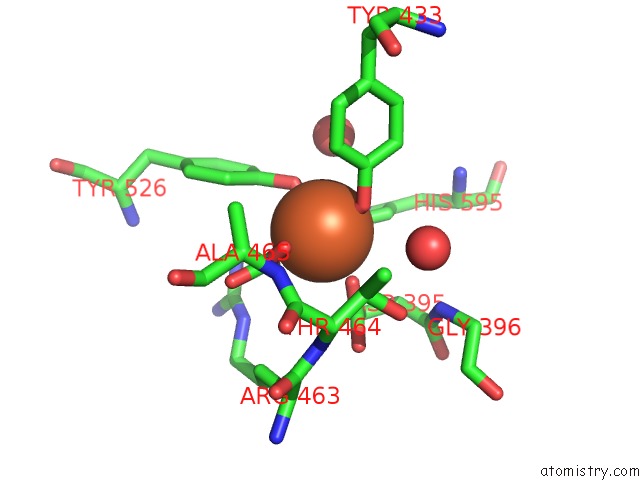
Mono view
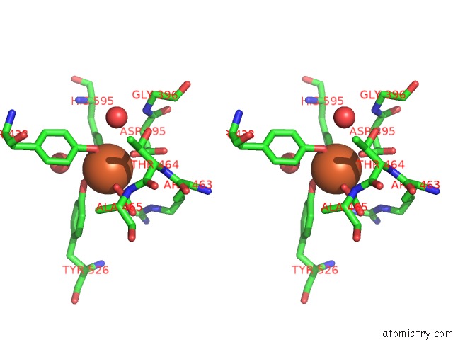
Stereo pair view
Mono view
Stereo pair view
A full contact list of Iron with other atoms in the Fe binding
site number 1 of Crystal Structure of C-Lobe of Bovine Lactoferrin Complexed with Aminocaproic Acid at 1.46 A Resolution within 5.0Å range:
|
Reference:
P.K.Shukla,
L.Gautam,
M.Sinha,
P.Kaur,
S.Sharma,
T.P.Singh.
Crystal Structure of C-Lobe of Bovine Lactoferrin Complexed with Aminocaproic Acid at 1.46 A Resolution To Be Published.
Page generated: Tue Aug 5 09:57:25 2025
Last articles
Mg in 5LCBMg in 5LCF
Mg in 5LBO
Mg in 5LCD
Mg in 5LC8
Mg in 5LAJ
Mg in 5LAI
Mg in 5LBA
Mg in 5LB5
Mg in 5LB3