Iron »
PDB 4fh7-4g38 »
4fyz »
Iron in PDB 4fyz: Crystal Structure of Nitrosyl Cytochrome P450CIN
Protein crystallography data
The structure of Crystal Structure of Nitrosyl Cytochrome P450CIN, PDB code: 4fyz
was solved by
Y.Madrona,
S.M.Tripathi,
H.Li,
T.L.Poulos,
with X-Ray Crystallography technique. A brief refinement statistics is given in the table below:
| Resolution Low / High (Å) | 36.01 / 2.32 |
| Space group | P 1 21 1 |
| Cell size a, b, c (Å), α, β, γ (°) | 64.176, 68.437, 104.047, 90.00, 95.79, 90.00 |
| R / Rfree (%) | 19.3 / 25.1 |
Iron Binding Sites:
The binding sites of Iron atom in the Crystal Structure of Nitrosyl Cytochrome P450CIN
(pdb code 4fyz). This binding sites where shown within
5.0 Angstroms radius around Iron atom.
In total 2 binding sites of Iron where determined in the Crystal Structure of Nitrosyl Cytochrome P450CIN, PDB code: 4fyz:
Jump to Iron binding site number: 1; 2;
In total 2 binding sites of Iron where determined in the Crystal Structure of Nitrosyl Cytochrome P450CIN, PDB code: 4fyz:
Jump to Iron binding site number: 1; 2;
Iron binding site 1 out of 2 in 4fyz
Go back to
Iron binding site 1 out
of 2 in the Crystal Structure of Nitrosyl Cytochrome P450CIN
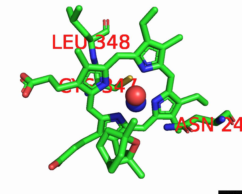
Mono view
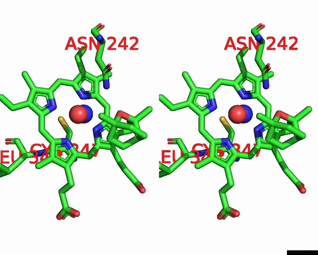
Stereo pair view
Mono view
Stereo pair view
A full contact list of Iron with other atoms in the Fe binding
site number 1 of Crystal Structure of Nitrosyl Cytochrome P450CIN within 5.0Å range:
|
Iron binding site 2 out of 2 in 4fyz
Go back to
Iron binding site 2 out
of 2 in the Crystal Structure of Nitrosyl Cytochrome P450CIN
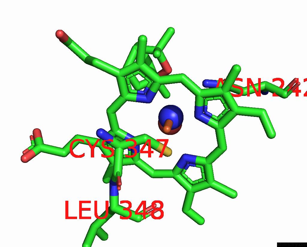
Mono view
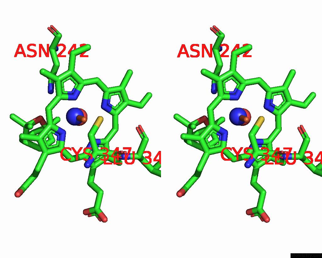
Stereo pair view
Mono view
Stereo pair view
A full contact list of Iron with other atoms in the Fe binding
site number 2 of Crystal Structure of Nitrosyl Cytochrome P450CIN within 5.0Å range:
|
Reference:
Y.Madrona,
S.Tripathi,
H.Li,
T.L.Poulos.
Crystal Structures of Substrate-Free and Nitrosyl Cytochrome P450CIN: Implications For O(2) Activation. Biochemistry V. 51 6623 2012.
ISSN: ISSN 0006-2960
PubMed: 22775403
DOI: 10.1021/BI300666U
Page generated: Tue Aug 5 10:31:13 2025
ISSN: ISSN 0006-2960
PubMed: 22775403
DOI: 10.1021/BI300666U
Last articles
Xe in 4ZZBXe in 4YLH
Xe in 3U52
Xe in 4WNA
Xe in 4WN9
Xe in 4O4T
Xe in 4NXA
Xe in 4PWB
Xe in 4GUV
Xe in 4NWH