Atomistry » Iron » PDB 4fh7-4g38 » 4g1x
Atomistry »
  Iron »
    PDB 4fh7-4g38 »
      4g1x »

Iron in PDB 4g1x: Crystal Structure of Mycobacterium Tuberculosis CYP121 in Complex with 4-(1H-1,2,4-Triazol-1-Yl)Quinolin-6-Amine

Protein crystallography data

The structure of Crystal Structure of Mycobacterium Tuberculosis CYP121 in Complex with 4-(1H-1,2,4-Triazol-1-Yl)Quinolin-6-Amine, PDB code: 4g1x was solved by S.A.Hudson, with X-Ray Crystallography technique. A brief refinement statistics is given in the table below:

Resolution Low / High (Å) 37.14 / 1.30
Space group P 65 2 2
Cell size a, b, c (Å), α, β, γ (°) 77.407, 77.407, 263.954, 90.00, 90.00, 120.00
R / Rfree (%) 16.2 / 18.6

Iron Binding Sites:

The binding sites of Iron atom in the Crystal Structure of Mycobacterium Tuberculosis CYP121 in Complex with 4-(1H-1,2,4-Triazol-1-Yl)Quinolin-6-Amine (pdb code 4g1x). This binding sites where shown within 5.0 Angstroms radius around Iron atom.
In total 2 binding sites of Iron where determined in the Crystal Structure of Mycobacterium Tuberculosis CYP121 in Complex with 4-(1H-1,2,4-Triazol-1-Yl)Quinolin-6-Amine, PDB code: 4g1x:
Jump to Iron binding site number: 1; 2;

Iron binding site 1 out of 2 in 4g1x

Go back to Iron Binding Sites List in 4g1x
Iron binding site 1 out of 2 in the Crystal Structure of Mycobacterium Tuberculosis CYP121 in Complex with 4-(1H-1,2,4-Triazol-1-Yl)Quinolin-6-Amine
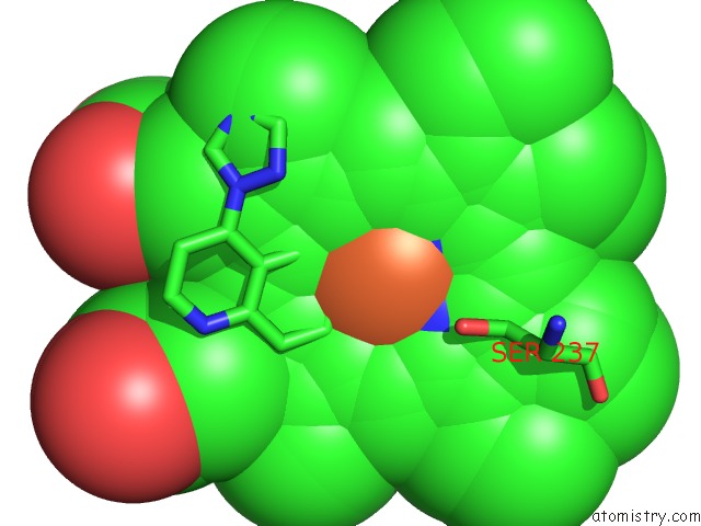

Mono view
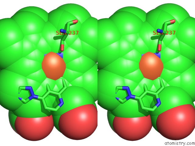

Stereo pair view

A full contact list of Iron with other atoms in the Fe binding site number 1 of Crystal Structure of Mycobacterium Tuberculosis CYP121 in Complex with 4-(1H-1,2,4-Triazol-1-Yl)Quinolin-6-Amine within 5.0Å range:
probe atom residue distance (Å) B Occ
A:Fe401

b:7.3
occ:0.70
FE A:HEM401 0.0 7.3 0.7
FE A:HEM401 0.3 4.5 0.3
NC A:HEM401 1.8 4.6 0.3
NA A:HEM401 1.9 6.9 0.7
ND A:HEM401 2.0 5.0 0.3
NC A:HEM401 2.0 7.5 0.7
NB A:HEM401 2.0 6.7 0.7
ND A:HEM401 2.0 8.9 0.7
NA A:HEM401 2.1 5.2 0.3
NB A:HEM401 2.2 4.8 0.3
SG A:CYS345 2.2 7.3 1.0
NAA A:TQU409 2.4 8.8 1.0
C4C A:HEM401 2.9 4.6 0.3
C1D A:HEM401 2.9 5.2 0.3
C1C A:HEM401 2.9 4.5 0.3
C1A A:HEM401 3.0 7.4 0.7
C1C A:HEM401 3.0 7.0 0.7
C4B A:HEM401 3.0 6.7 0.7
C4D A:HEM401 3.0 5.3 0.3
C1D A:HEM401 3.0 9.3 0.7
C1B A:HEM401 3.0 6.2 0.7
C4D A:HEM401 3.1 8.9 0.7
C4C A:HEM401 3.1 7.9 0.7
C4A A:HEM401 3.1 6.6 0.7
C4B A:HEM401 3.1 4.8 0.3
C1A A:HEM401 3.1 5.2 0.3
CAL A:TQU409 3.2 7.5 1.0
CHD A:HEM401 3.2 4.6 0.3
C4A A:HEM401 3.2 5.2 0.3
C1B A:HEM401 3.3 4.9 0.3
CB A:CYS345 3.3 8.0 1.0
CHD A:HEM401 3.4 8.9 0.7
CHA A:HEM401 3.4 7.9 0.7
CHC A:HEM401 3.4 4.8 0.3
CHB A:HEM401 3.5 6.1 0.7
CHC A:HEM401 3.5 7.4 0.7
CHA A:HEM401 3.5 5.1 0.3
CAD A:TQU409 3.7 8.4 1.0
CHB A:HEM401 3.7 5.5 0.3
CAH A:TQU409 4.1 9.3 1.0
CA A:CYS345 4.1 7.2 1.0
C3C A:HEM401 4.1 4.7 0.3
C2C A:HEM401 4.2 4.5 0.3
C2D A:HEM401 4.2 5.2 0.3
C3D A:HEM401 4.2 5.6 0.3
C2B A:HEM401 4.2 7.0 0.7
C2A A:HEM401 4.3 7.2 0.7
C2C A:HEM401 4.3 8.8 0.7
C3C A:HEM401 4.3 8.2 0.7
C3B A:HEM401 4.3 6.6 0.7
C3A A:HEM401 4.3 6.5 0.7
C3D A:HEM401 4.3 9.8 0.7
C2D A:HEM401 4.3 10.2 0.7
C2A A:HEM401 4.4 5.2 0.3
C3B A:HEM401 4.4 5.2 0.3
C3A A:HEM401 4.4 5.3 0.3
C2B A:HEM401 4.5 5.2 0.3
OG A:SER237 4.7 6.4 1.0
N A:GLY347 4.9 8.3 1.0
CAF A:TQU409 4.9 9.0 1.0
C A:CYS345 4.9 7.9 1.0
CD A:PRO346 4.9 9.0 1.0

Iron binding site 2 out of 2 in 4g1x

Go back to Iron Binding Sites List in 4g1x
Iron binding site 2 out of 2 in the Crystal Structure of Mycobacterium Tuberculosis CYP121 in Complex with 4-(1H-1,2,4-Triazol-1-Yl)Quinolin-6-Amine
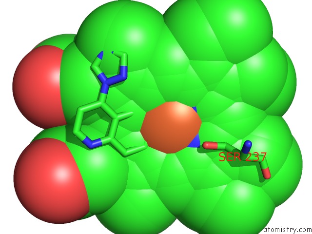

Mono view
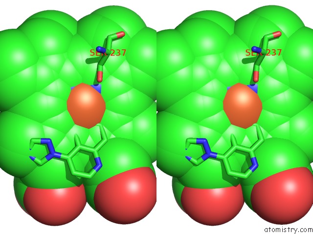

Stereo pair view

A full contact list of Iron with other atoms in the Fe binding site number 2 of Crystal Structure of Mycobacterium Tuberculosis CYP121 in Complex with 4-(1H-1,2,4-Triazol-1-Yl)Quinolin-6-Amine within 5.0Å range:
probe atom residue distance (Å) B Occ
A:Fe401

b:4.5
occ:0.30
FE A:HEM401 0.0 4.5 0.3
FE A:HEM401 0.3 7.3 0.7
NC A:HEM401 1.9 7.5 0.7
ND A:HEM401 1.9 8.9 0.7
NA A:HEM401 1.9 5.2 0.3
NC A:HEM401 1.9 4.6 0.3
NA A:HEM401 2.1 6.9 0.7
NB A:HEM401 2.1 4.8 0.3
ND A:HEM401 2.1 5.0 0.3
NB A:HEM401 2.2 6.7 0.7
NAA A:TQU409 2.2 8.8 1.0
SG A:CYS345 2.4 7.3 1.0
C1D A:HEM401 2.8 9.3 0.7
C4C A:HEM401 2.9 7.9 0.7
C4D A:HEM401 3.0 8.9 0.7
C1C A:HEM401 3.0 7.0 0.7
C1A A:HEM401 3.0 5.2 0.3
CAL A:TQU409 3.0 7.5 1.0
C4C A:HEM401 3.0 4.6 0.3
C4B A:HEM401 3.0 4.8 0.3
C4A A:HEM401 3.0 5.2 0.3
C1C A:HEM401 3.0 4.5 0.3
C1D A:HEM401 3.0 5.2 0.3
C1A A:HEM401 3.1 7.4 0.7
C4D A:HEM401 3.1 5.3 0.3
C1B A:HEM401 3.1 4.9 0.3
C4B A:HEM401 3.1 6.7 0.7
CHD A:HEM401 3.2 8.9 0.7
C1B A:HEM401 3.2 6.2 0.7
C4A A:HEM401 3.2 6.6 0.7
CHD A:HEM401 3.4 4.6 0.3
CHA A:HEM401 3.4 7.9 0.7
CHC A:HEM401 3.5 4.8 0.3
CHA A:HEM401 3.5 5.1 0.3
CB A:CYS345 3.5 8.0 1.0
CHC A:HEM401 3.5 7.4 0.7
CHB A:HEM401 3.5 5.5 0.3
CAD A:TQU409 3.6 8.4 1.0
CHB A:HEM401 3.6 6.1 0.7
CAH A:TQU409 3.9 9.3 1.0
C3C A:HEM401 4.1 8.2 0.7
C2D A:HEM401 4.2 10.2 0.7
C3D A:HEM401 4.2 9.8 0.7
C2C A:HEM401 4.2 8.8 0.7
CA A:CYS345 4.2 7.2 1.0
C2A A:HEM401 4.2 5.2 0.3
C3A A:HEM401 4.3 5.3 0.3
C3C A:HEM401 4.3 4.7 0.3
C3B A:HEM401 4.3 5.2 0.3
C2C A:HEM401 4.3 4.5 0.3
C2D A:HEM401 4.3 5.2 0.3
C2B A:HEM401 4.3 5.2 0.3
C3D A:HEM401 4.3 5.6 0.3
C2A A:HEM401 4.3 7.2 0.7
C3B A:HEM401 4.4 6.6 0.7
C2B A:HEM401 4.4 7.0 0.7
C3A A:HEM401 4.4 6.5 0.7
OG A:SER237 4.6 6.4 1.0
CAF A:TQU409 4.8 9.0 1.0
N A:GLY347 4.9 8.3 1.0
CD A:PRO346 4.9 9.0 1.0
CAO A:TQU409 5.0 10.2 1.0
C A:CYS345 5.0 7.9 1.0
CB A:SER237 5.0 6.0 1.0

Reference:

S.A.Hudson, K.J.Mclean, S.Surade, Y.-Q.Yang, D.Leys, A.Ciulli, A.W.Munro, C.Abell. Application of Fragment Screening and Merging to the Discovery of Inhibitors of the Mycobacterium Tuberculosis Cytochrome P450 CYP121 Angew.Chem.Int.Ed.Engl. V. 51 9311 2012.
ISSN: ISSN 1433-7851
PubMed: 22890978
DOI: 10.1002/ANIE.201202544
Page generated: Tue Aug 5 10:31:46 2025

Last articles

Zn in 9UUO
Zn in 9UUS
Zn in 9W4R
Zn in 9VKW
Zn in 9W4S
Zn in 9VH1
Zn in 9RMX
Zn in 9RMU
Zn in 9QWN
Zn in 9U9Y
© Copyright 2008-2020 by atomistry.com
Home   |    Site Map   |    Copyright   |    Contact us   |    Privacy