Iron »
PDB 4gl7-4h9l »
4h1n »
Iron in PDB 4h1n: Crystal Structure of P450 2B4 F297A Mutant in Complex with Anti- Platelet Drug Clopidogrel
Enzymatic activity of Crystal Structure of P450 2B4 F297A Mutant in Complex with Anti- Platelet Drug Clopidogrel
All present enzymatic activity of Crystal Structure of P450 2B4 F297A Mutant in Complex with Anti- Platelet Drug Clopidogrel:
1.14.14.1;
1.14.14.1;
Protein crystallography data
The structure of Crystal Structure of P450 2B4 F297A Mutant in Complex with Anti- Platelet Drug Clopidogrel, PDB code: 4h1n
was solved by
M.B.Shah,
H.H.Jang,
C.D.Stout,
J.R.Halpert,
with X-Ray Crystallography technique. A brief refinement statistics is given in the table below:
| Resolution Low / High (Å) | 20.00 / 2.99 |
| Space group | P 31 2 1 |
| Cell size a, b, c (Å), α, β, γ (°) | 91.385, 91.385, 152.286, 90.00, 90.00, 120.00 |
| R / Rfree (%) | 23.3 / 28.6 |
Other elements in 4h1n:
The structure of Crystal Structure of P450 2B4 F297A Mutant in Complex with Anti- Platelet Drug Clopidogrel also contains other interesting chemical elements:
| Chlorine | (Cl) | 1 atom |
Iron Binding Sites:
The binding sites of Iron atom in the Crystal Structure of P450 2B4 F297A Mutant in Complex with Anti- Platelet Drug Clopidogrel
(pdb code 4h1n). This binding sites where shown within
5.0 Angstroms radius around Iron atom.
In total only one binding site of Iron was determined in the Crystal Structure of P450 2B4 F297A Mutant in Complex with Anti- Platelet Drug Clopidogrel, PDB code: 4h1n:
In total only one binding site of Iron was determined in the Crystal Structure of P450 2B4 F297A Mutant in Complex with Anti- Platelet Drug Clopidogrel, PDB code: 4h1n:
Iron binding site 1 out of 1 in 4h1n
Go back to
Iron binding site 1 out
of 1 in the Crystal Structure of P450 2B4 F297A Mutant in Complex with Anti- Platelet Drug Clopidogrel

Mono view
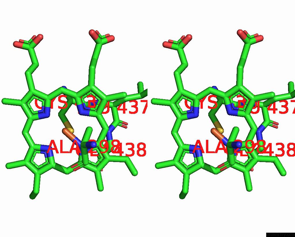
Stereo pair view
Mono view
Stereo pair view
A full contact list of Iron with other atoms in the Fe binding
site number 1 of Crystal Structure of P450 2B4 F297A Mutant in Complex with Anti- Platelet Drug Clopidogrel within 5.0Å range:
|
Reference:
M.B.Shah,
H.H.Jang,
Q.Zhang,
C.David Stout,
J.R.Halpert.
X-Ray Crystal Structure of the Cytochrome P450 2B4 Active Site Mutant F297A in Complex with Clopidogrel: Insights Into Compensatory Rearrangements of the Binding Pocket. Arch.Biochem.Biophys. V. 530 64 2013.
ISSN: ISSN 0003-9861
PubMed: 23296089
DOI: 10.1016/J.ABB.2012.12.016
Page generated: Mon Aug 5 03:07:41 2024
ISSN: ISSN 0003-9861
PubMed: 23296089
DOI: 10.1016/J.ABB.2012.12.016
Last articles
Cl in 8A6QCl in 8A6O
Cl in 8A2C
Cl in 8A6N
Cl in 8A6G
Cl in 8A6F
Cl in 8A6H
Cl in 8A67
Cl in 8A62
Cl in 8A65