Iron »
PDB 4h9t-4hm6 »
4hhr »
Iron in PDB 4hhr: Crystal Structure of Fatty Acid Alpha-Dioxygenase (Arabidopsis Thaliana)
Protein crystallography data
The structure of Crystal Structure of Fatty Acid Alpha-Dioxygenase (Arabidopsis Thaliana), PDB code: 4hhr
was solved by
C.C.Goulah,
G.Zhu,
M.Koszelak-Rosenblum,
M.G.Malkowski,
with X-Ray Crystallography technique. A brief refinement statistics is given in the table below:
| Resolution Low / High (Å) | 47.78 / 1.51 |
| Space group | P 43 2 2 |
| Cell size a, b, c (Å), α, β, γ (°) | 101.935, 101.935, 137.510, 90.00, 90.00, 90.00 |
| R / Rfree (%) | 15.3 / 18.2 |
Other elements in 4hhr:
The structure of Crystal Structure of Fatty Acid Alpha-Dioxygenase (Arabidopsis Thaliana) also contains other interesting chemical elements:
| Chlorine | (Cl) | 3 atoms |
| Calcium | (Ca) | 8 atoms |
Iron Binding Sites:
The binding sites of Iron atom in the Crystal Structure of Fatty Acid Alpha-Dioxygenase (Arabidopsis Thaliana)
(pdb code 4hhr). This binding sites where shown within
5.0 Angstroms radius around Iron atom.
In total only one binding site of Iron was determined in the Crystal Structure of Fatty Acid Alpha-Dioxygenase (Arabidopsis Thaliana), PDB code: 4hhr:
In total only one binding site of Iron was determined in the Crystal Structure of Fatty Acid Alpha-Dioxygenase (Arabidopsis Thaliana), PDB code: 4hhr:
Iron binding site 1 out of 1 in 4hhr
Go back to
Iron binding site 1 out
of 1 in the Crystal Structure of Fatty Acid Alpha-Dioxygenase (Arabidopsis Thaliana)
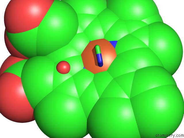
Mono view
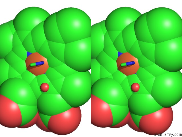
Stereo pair view
Mono view
Stereo pair view
A full contact list of Iron with other atoms in the Fe binding
site number 1 of Crystal Structure of Fatty Acid Alpha-Dioxygenase (Arabidopsis Thaliana) within 5.0Å range:
|
Reference:
C.C.Goulah,
G.Zhu,
M.Koszelak-Rosenblum,
M.G.Malkowski.
The Crystal Structure of Alpha-Dioxygenase Provides Insight Into Diversity in the Cyclooxygenase-Peroxidase Superfamily. Biochemistry V. 52 1364 2013.
ISSN: ISSN 0006-2960
PubMed: 23373518
DOI: 10.1021/BI400013K
Page generated: Tue Aug 5 10:57:15 2025
ISSN: ISSN 0006-2960
PubMed: 23373518
DOI: 10.1021/BI400013K
Last articles
Mg in 5N78Mg in 5N77
Mg in 5N6Y
Mg in 5N75
Mg in 5N6O
Mg in 5N5N
Mg in 5N69
Mg in 5N6A
Mg in 5N61
Mg in 5N5Y