Iron »
PDB 4hm7-4ies »
4hsx »
Iron in PDB 4hsx: Structure of the L100F Mutant of Dehaloperoxidase-Hemoglobin A From Amphitrite Ornata with 4-Bromophenol
Enzymatic activity of Structure of the L100F Mutant of Dehaloperoxidase-Hemoglobin A From Amphitrite Ornata with 4-Bromophenol
All present enzymatic activity of Structure of the L100F Mutant of Dehaloperoxidase-Hemoglobin A From Amphitrite Ornata with 4-Bromophenol:
1.11.1.7;
1.11.1.7;
Protein crystallography data
The structure of Structure of the L100F Mutant of Dehaloperoxidase-Hemoglobin A From Amphitrite Ornata with 4-Bromophenol, PDB code: 4hsx
was solved by
M.K.Thompson,
A.Plummer,
S.Franzen,
with X-Ray Crystallography technique. A brief refinement statistics is given in the table below:
| Resolution Low / High (Å) | 48.34 / 1.12 |
| Space group | P 21 21 21 |
| Cell size a, b, c (Å), α, β, γ (°) | 58.241, 67.441, 69.338, 90.00, 90.00, 90.00 |
| R / Rfree (%) | 12.6 / 15 |
Other elements in 4hsx:
The structure of Structure of the L100F Mutant of Dehaloperoxidase-Hemoglobin A From Amphitrite Ornata with 4-Bromophenol also contains other interesting chemical elements:
| Bromine | (Br) | 2 atoms |
Iron Binding Sites:
The binding sites of Iron atom in the Structure of the L100F Mutant of Dehaloperoxidase-Hemoglobin A From Amphitrite Ornata with 4-Bromophenol
(pdb code 4hsx). This binding sites where shown within
5.0 Angstroms radius around Iron atom.
In total 2 binding sites of Iron where determined in the Structure of the L100F Mutant of Dehaloperoxidase-Hemoglobin A From Amphitrite Ornata with 4-Bromophenol, PDB code: 4hsx:
Jump to Iron binding site number: 1; 2;
In total 2 binding sites of Iron where determined in the Structure of the L100F Mutant of Dehaloperoxidase-Hemoglobin A From Amphitrite Ornata with 4-Bromophenol, PDB code: 4hsx:
Jump to Iron binding site number: 1; 2;
Iron binding site 1 out of 2 in 4hsx
Go back to
Iron binding site 1 out
of 2 in the Structure of the L100F Mutant of Dehaloperoxidase-Hemoglobin A From Amphitrite Ornata with 4-Bromophenol
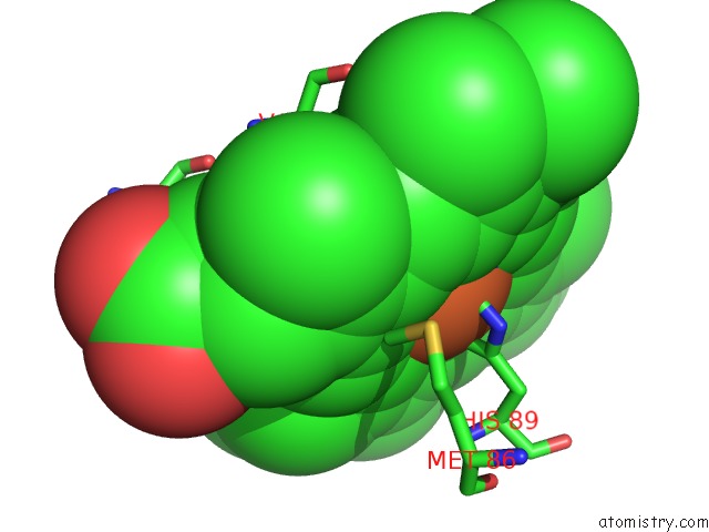
Mono view
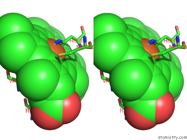
Stereo pair view
Mono view
Stereo pair view
A full contact list of Iron with other atoms in the Fe binding
site number 1 of Structure of the L100F Mutant of Dehaloperoxidase-Hemoglobin A From Amphitrite Ornata with 4-Bromophenol within 5.0Å range:
|
Iron binding site 2 out of 2 in 4hsx
Go back to
Iron binding site 2 out
of 2 in the Structure of the L100F Mutant of Dehaloperoxidase-Hemoglobin A From Amphitrite Ornata with 4-Bromophenol
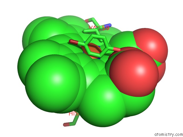
Mono view

Stereo pair view
Mono view
Stereo pair view
A full contact list of Iron with other atoms in the Fe binding
site number 2 of Structure of the L100F Mutant of Dehaloperoxidase-Hemoglobin A From Amphitrite Ornata with 4-Bromophenol within 5.0Å range:
|
Reference:
A.Plummer,
M.K.Thompson,
S.Franzen.
Role of Polarity of the Distal Pocket in the Control of Inhibitor Binding in Dehaloperoxidase-Hemoglobin. Biochemistry V. 52 2218 2013.
ISSN: ISSN 0006-2960
PubMed: 23480794
DOI: 10.1021/BI301509R
Page generated: Tue Aug 5 11:09:24 2025
ISSN: ISSN 0006-2960
PubMed: 23480794
DOI: 10.1021/BI301509R
Last articles
Mg in 6IK9Mg in 6IJI
Mg in 6IJH
Mg in 6IID
Mg in 6III
Mg in 6IHW
Mg in 6II6
Mg in 6IHV
Mg in 6IHU
Mg in 6IHS