Iron »
PDB 4iet-4ixq »
4itk »
Iron in PDB 4itk: The Structure of C.Reinhardtii Ferredoxin 2
Protein crystallography data
The structure of The Structure of C.Reinhardtii Ferredoxin 2, PDB code: 4itk
was solved by
P.M.Alahuhta,
V.V.Lunin,
with X-Ray Crystallography technique. A brief refinement statistics is given in the table below:
| Resolution Low / High (Å) | 25.00 / 1.18 |
| Space group | P 1 |
| Cell size a, b, c (Å), α, β, γ (°) | 25.429, 26.458, 31.016, 102.56, 104.35, 100.30 |
| R / Rfree (%) | 10.9 / 14.7 |
Iron Binding Sites:
The binding sites of Iron atom in the The Structure of C.Reinhardtii Ferredoxin 2
(pdb code 4itk). This binding sites where shown within
5.0 Angstroms radius around Iron atom.
In total 2 binding sites of Iron where determined in the The Structure of C.Reinhardtii Ferredoxin 2, PDB code: 4itk:
Jump to Iron binding site number: 1; 2;
In total 2 binding sites of Iron where determined in the The Structure of C.Reinhardtii Ferredoxin 2, PDB code: 4itk:
Jump to Iron binding site number: 1; 2;
Iron binding site 1 out of 2 in 4itk
Go back to
Iron binding site 1 out
of 2 in the The Structure of C.Reinhardtii Ferredoxin 2
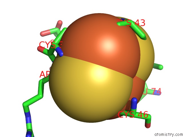
Mono view
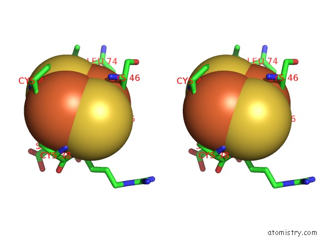
Stereo pair view
Mono view
Stereo pair view
A full contact list of Iron with other atoms in the Fe binding
site number 1 of The Structure of C.Reinhardtii Ferredoxin 2 within 5.0Å range:
|
Iron binding site 2 out of 2 in 4itk
Go back to
Iron binding site 2 out
of 2 in the The Structure of C.Reinhardtii Ferredoxin 2
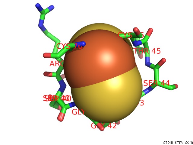
Mono view
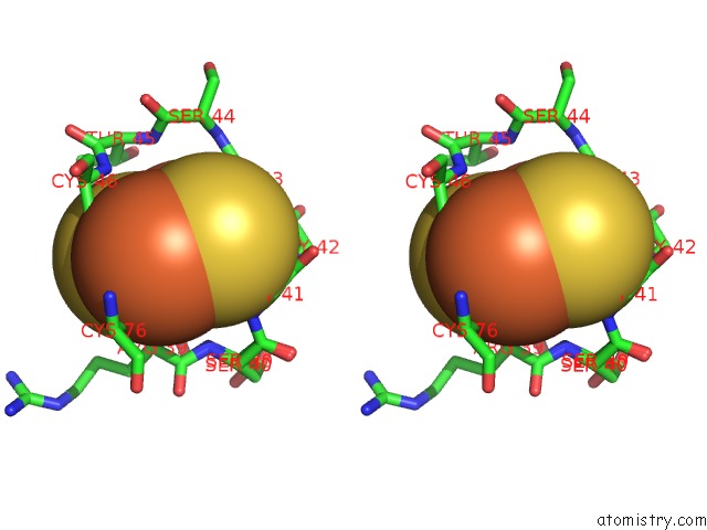
Stereo pair view
Mono view
Stereo pair view
A full contact list of Iron with other atoms in the Fe binding
site number 2 of The Structure of C.Reinhardtii Ferredoxin 2 within 5.0Å range:
|
Reference:
P.M.Alahuhta,
V.V.Lunin.
C.Reinhardtii Ferredoxin 2 To Be Published.
Page generated: Tue Aug 5 11:20:12 2025
Last articles
Mg in 3I2BMg in 3I33
Mg in 3HZY
Mg in 3HZV
Mg in 3HZM
Mg in 3I0O
Mg in 3HZT
Mg in 3HZK
Mg in 3HYT
Mg in 3HY6