Iron »
PDB 4ixr-4jn0 »
4jlt »
Iron in PDB 4jlt: Crystal Structure of P450 2B4(H226Y) in Complex with Paroxetine
Enzymatic activity of Crystal Structure of P450 2B4(H226Y) in Complex with Paroxetine
All present enzymatic activity of Crystal Structure of P450 2B4(H226Y) in Complex with Paroxetine:
1.14.14.1;
1.14.14.1;
Protein crystallography data
The structure of Crystal Structure of P450 2B4(H226Y) in Complex with Paroxetine, PDB code: 4jlt
was solved by
M.B.Shah,
J.Pascual,
C.D.Stout,
J.R.Halpert,
with X-Ray Crystallography technique. A brief refinement statistics is given in the table below:
| Resolution Low / High (Å) | 78.60 / 2.14 |
| Space group | P 31 2 1 |
| Cell size a, b, c (Å), α, β, γ (°) | 90.760, 90.760, 153.681, 90.00, 90.00, 120.00 |
| R / Rfree (%) | 18.7 / 22.9 |
Other elements in 4jlt:
The structure of Crystal Structure of P450 2B4(H226Y) in Complex with Paroxetine also contains other interesting chemical elements:
| Fluorine | (F) | 1 atom |
Iron Binding Sites:
The binding sites of Iron atom in the Crystal Structure of P450 2B4(H226Y) in Complex with Paroxetine
(pdb code 4jlt). This binding sites where shown within
5.0 Angstroms radius around Iron atom.
In total only one binding site of Iron was determined in the Crystal Structure of P450 2B4(H226Y) in Complex with Paroxetine, PDB code: 4jlt:
In total only one binding site of Iron was determined in the Crystal Structure of P450 2B4(H226Y) in Complex with Paroxetine, PDB code: 4jlt:
Iron binding site 1 out of 1 in 4jlt
Go back to
Iron binding site 1 out
of 1 in the Crystal Structure of P450 2B4(H226Y) in Complex with Paroxetine
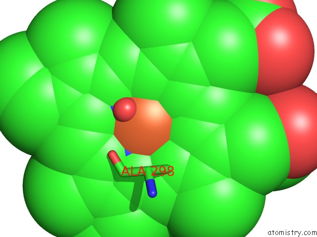
Mono view
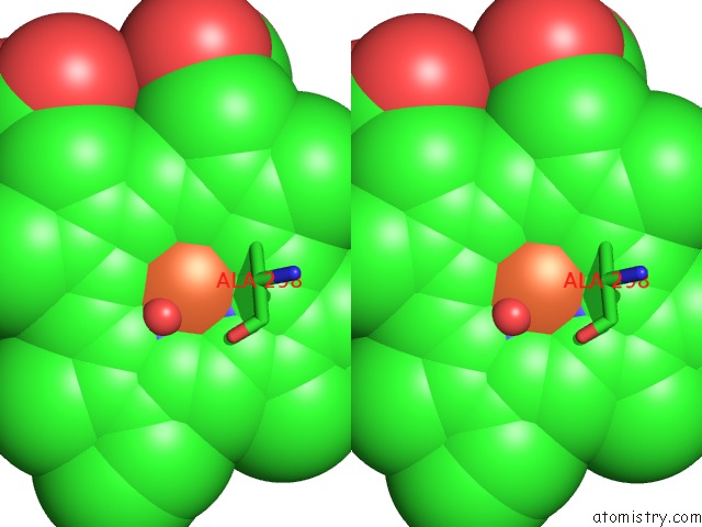
Stereo pair view
Mono view
Stereo pair view
A full contact list of Iron with other atoms in the Fe binding
site number 1 of Crystal Structure of P450 2B4(H226Y) in Complex with Paroxetine within 5.0Å range:
|
Reference:
M.B.Shah,
I.Kufareva,
J.Pascual,
Q.Zhang,
C.D.Stout,
J.R.Halpert.
A Structural Snapshot of CYP2B4 in Complex with Paroxetine Provides Insights Into Ligand Binding and Clusters of Conformational States. J.Pharmacol.Exp.Ther. V. 346 113 2013.
ISSN: ISSN 0022-3565
PubMed: 23633618
DOI: 10.1124/JPET.113.204776
Page generated: Tue Aug 5 11:35:39 2025
ISSN: ISSN 0022-3565
PubMed: 23633618
DOI: 10.1124/JPET.113.204776
Last articles
Mg in 6CT2Mg in 6CRC
Mg in 6CR9
Mg in 6CRB
Mg in 6CR8
Mg in 6CR7
Mg in 6CR5
Mg in 6CR4
Mg in 6CR6
Mg in 6CR3