Iron »
PDB 4m71-4n0k »
4mhu »
Iron in PDB 4mhu: Crystal Structure of Ectd From S. Alaskensis with Bound Fe
Protein crystallography data
The structure of Crystal Structure of Ectd From S. Alaskensis with Bound Fe, PDB code: 4mhu
was solved by
N.Widderich,
A.Hoeppner,
M.Pittelkow,
J.Heider,
S.H.Smits,
E.Bremer,
with X-Ray Crystallography technique. A brief refinement statistics is given in the table below:
| Resolution Low / High (Å) | 46.00 / 2.56 |
| Space group | P 21 21 21 |
| Cell size a, b, c (Å), α, β, γ (°) | 78.165, 87.519, 96.045, 90.00, 90.00, 90.00 |
| R / Rfree (%) | 20.2 / 26.6 |
Iron Binding Sites:
The binding sites of Iron atom in the Crystal Structure of Ectd From S. Alaskensis with Bound Fe
(pdb code 4mhu). This binding sites where shown within
5.0 Angstroms radius around Iron atom.
In total 2 binding sites of Iron where determined in the Crystal Structure of Ectd From S. Alaskensis with Bound Fe, PDB code: 4mhu:
Jump to Iron binding site number: 1; 2;
In total 2 binding sites of Iron where determined in the Crystal Structure of Ectd From S. Alaskensis with Bound Fe, PDB code: 4mhu:
Jump to Iron binding site number: 1; 2;
Iron binding site 1 out of 2 in 4mhu
Go back to
Iron binding site 1 out
of 2 in the Crystal Structure of Ectd From S. Alaskensis with Bound Fe
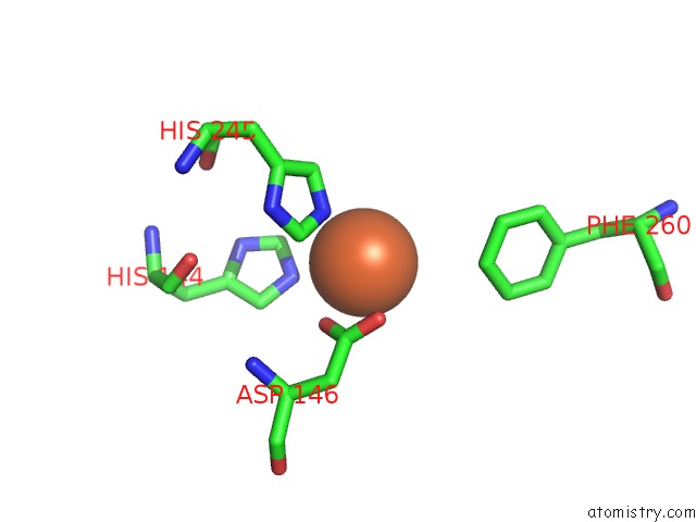
Mono view
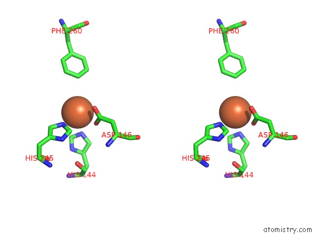
Stereo pair view
Mono view
Stereo pair view
A full contact list of Iron with other atoms in the Fe binding
site number 1 of Crystal Structure of Ectd From S. Alaskensis with Bound Fe within 5.0Å range:
|
Iron binding site 2 out of 2 in 4mhu
Go back to
Iron binding site 2 out
of 2 in the Crystal Structure of Ectd From S. Alaskensis with Bound Fe
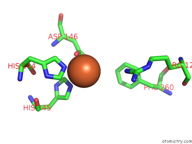
Mono view
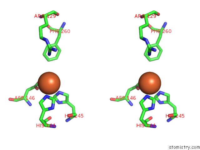
Stereo pair view
Mono view
Stereo pair view
A full contact list of Iron with other atoms in the Fe binding
site number 2 of Crystal Structure of Ectd From S. Alaskensis with Bound Fe within 5.0Å range:
|
Reference:
A.Hoppner,
N.Widderich,
M.Lenders,
E.Bremer,
S.H.Smits.
Crystal Structure of the Ectoine Hydroxylase, A Snapshot of the Active Site. J.Biol.Chem. V. 289 29570 2014.
ISSN: ISSN 0021-9258
PubMed: 25172507
DOI: 10.1074/JBC.M114.576769
Page generated: Tue Aug 5 12:47:05 2025
ISSN: ISSN 0021-9258
PubMed: 25172507
DOI: 10.1074/JBC.M114.576769
Last articles
Mn in 8ZWDMn in 8ZX9
Mn in 8ZX8
Mn in 8ZX2
Mn in 8ZX3
Mn in 8ZWY
Mn in 8ZWW
Mn in 8ZRL
Mn in 8ZWR
Mn in 8ZWP