Iron »
PDB 4n4j-4nji »
4n71 »
Iron in PDB 4n71: X-Ray Crystal Structure of 2-Amino-1-Hydroxyethylphosphonate-Bound Phnz
Protein crystallography data
The structure of X-Ray Crystal Structure of 2-Amino-1-Hydroxyethylphosphonate-Bound Phnz, PDB code: 4n71
was solved by
B.Woersdoerfer,
M.Lingaraju,
N.Yennawar,
A.K.Boal,
C.Krebs,
J.M.Bollingerjr,
M.-E.Pandelia,
with X-Ray Crystallography technique. A brief refinement statistics is given in the table below:
| Resolution Low / High (Å) | 29.09 / 2.98 |
| Space group | C 1 2 1 |
| Cell size a, b, c (Å), α, β, γ (°) | 148.761, 65.423, 109.355, 90.00, 124.83, 90.00 |
| R / Rfree (%) | 27.8 / 33.3 |
Iron Binding Sites:
The binding sites of Iron atom in the X-Ray Crystal Structure of 2-Amino-1-Hydroxyethylphosphonate-Bound Phnz
(pdb code 4n71). This binding sites where shown within
5.0 Angstroms radius around Iron atom.
In total 8 binding sites of Iron where determined in the X-Ray Crystal Structure of 2-Amino-1-Hydroxyethylphosphonate-Bound Phnz, PDB code: 4n71:
Jump to Iron binding site number: 1; 2; 3; 4; 5; 6; 7; 8;
In total 8 binding sites of Iron where determined in the X-Ray Crystal Structure of 2-Amino-1-Hydroxyethylphosphonate-Bound Phnz, PDB code: 4n71:
Jump to Iron binding site number: 1; 2; 3; 4; 5; 6; 7; 8;
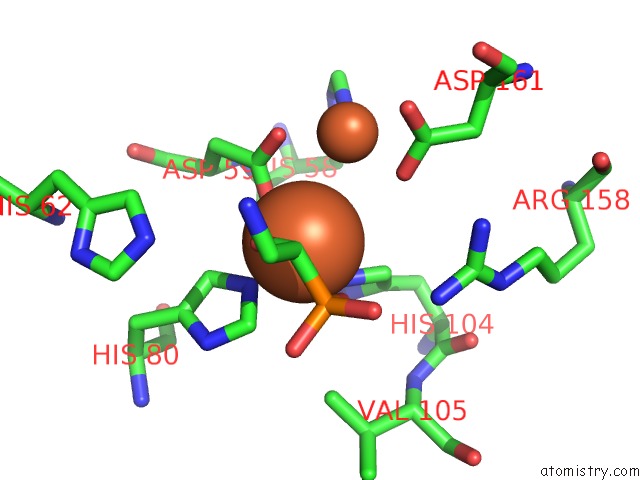
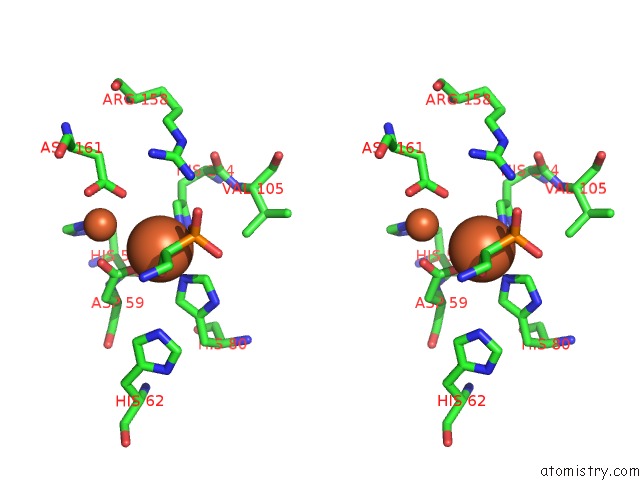
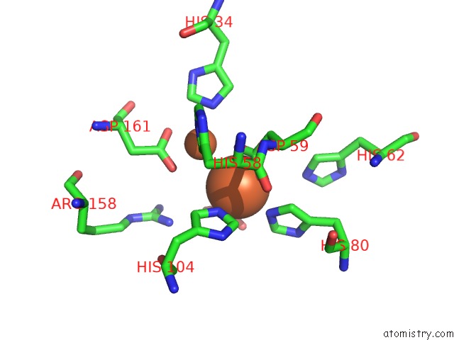
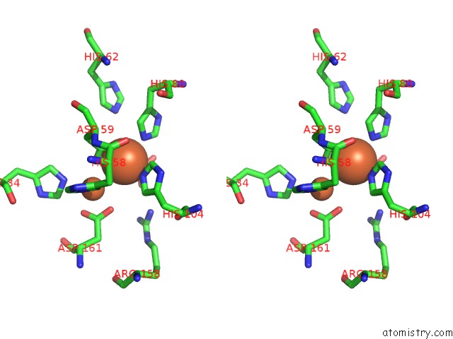
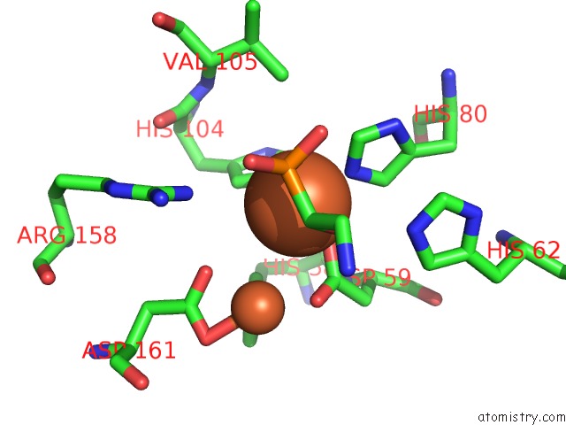
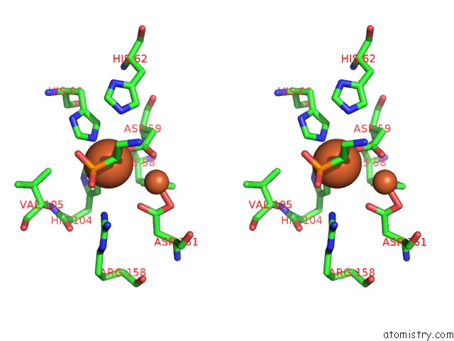
Iron binding site 1 out of 8 in 4n71
Go back to
Iron binding site 1 out
of 8 in the X-Ray Crystal Structure of 2-Amino-1-Hydroxyethylphosphonate-Bound Phnz
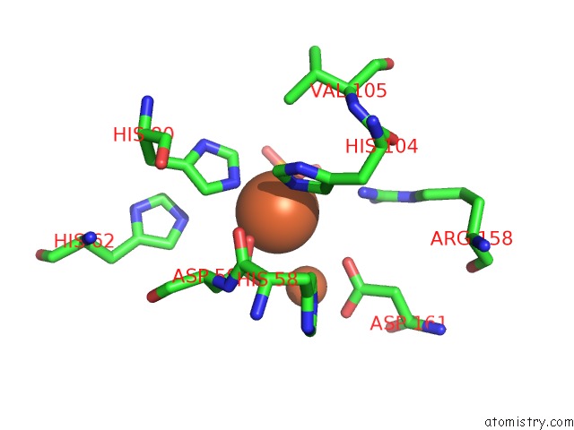
Mono view
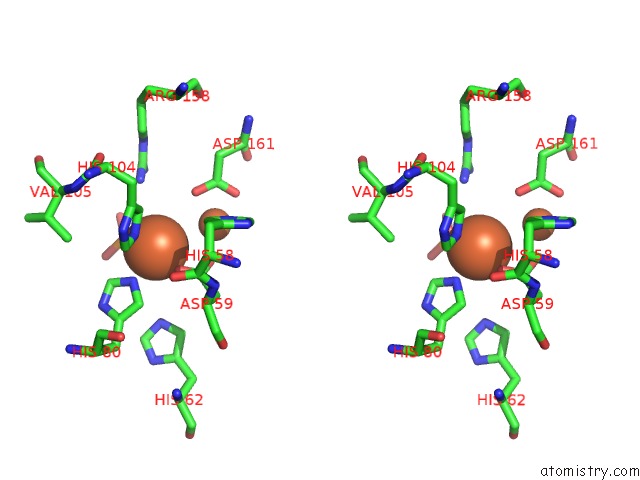
Stereo pair view
Mono view
Stereo pair view
A full contact list of Iron with other atoms in the Fe binding
site number 1 of X-Ray Crystal Structure of 2-Amino-1-Hydroxyethylphosphonate-Bound Phnz within 5.0Å range:
|
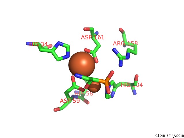
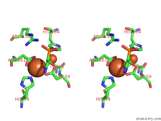
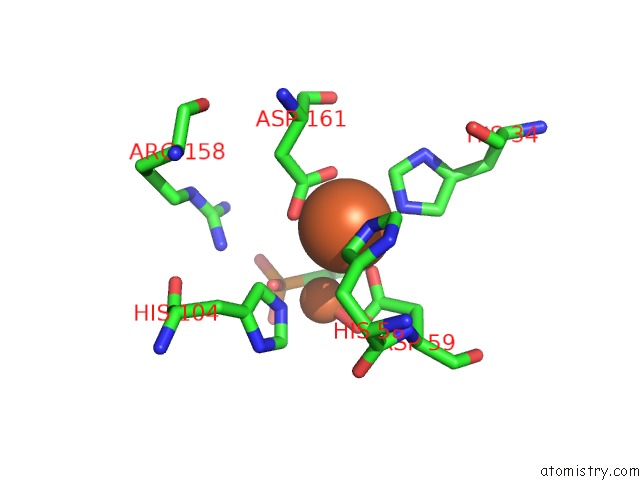
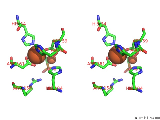
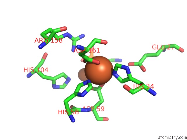
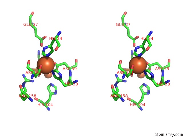
Iron binding site 2 out of 8 in 4n71
Go back to
Iron binding site 2 out
of 8 in the X-Ray Crystal Structure of 2-Amino-1-Hydroxyethylphosphonate-Bound Phnz
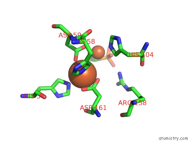
Mono view
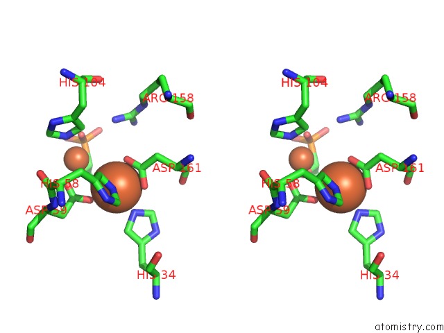
Stereo pair view
Mono view
Stereo pair view
A full contact list of Iron with other atoms in the Fe binding
site number 2 of X-Ray Crystal Structure of 2-Amino-1-Hydroxyethylphosphonate-Bound Phnz within 5.0Å range:
|
Iron binding site 3 out of 8 in 4n71
Go back to
Iron binding site 3 out
of 8 in the X-Ray Crystal Structure of 2-Amino-1-Hydroxyethylphosphonate-Bound Phnz
Mono view
Stereo pair view
Mono view
Stereo pair view
A full contact list of Iron with other atoms in the Fe binding
site number 3 of X-Ray Crystal Structure of 2-Amino-1-Hydroxyethylphosphonate-Bound Phnz within 5.0Å range:
|
Iron binding site 4 out of 8 in 4n71
Go back to
Iron binding site 4 out
of 8 in the X-Ray Crystal Structure of 2-Amino-1-Hydroxyethylphosphonate-Bound Phnz
Mono view
Stereo pair view
Mono view
Stereo pair view
A full contact list of Iron with other atoms in the Fe binding
site number 4 of X-Ray Crystal Structure of 2-Amino-1-Hydroxyethylphosphonate-Bound Phnz within 5.0Å range:
|
Iron binding site 5 out of 8 in 4n71
Go back to
Iron binding site 5 out
of 8 in the X-Ray Crystal Structure of 2-Amino-1-Hydroxyethylphosphonate-Bound Phnz
Mono view
Stereo pair view
Mono view
Stereo pair view
A full contact list of Iron with other atoms in the Fe binding
site number 5 of X-Ray Crystal Structure of 2-Amino-1-Hydroxyethylphosphonate-Bound Phnz within 5.0Å range:
|
Iron binding site 6 out of 8 in 4n71
Go back to
Iron binding site 6 out
of 8 in the X-Ray Crystal Structure of 2-Amino-1-Hydroxyethylphosphonate-Bound Phnz
Mono view
Stereo pair view
Mono view
Stereo pair view
A full contact list of Iron with other atoms in the Fe binding
site number 6 of X-Ray Crystal Structure of 2-Amino-1-Hydroxyethylphosphonate-Bound Phnz within 5.0Å range:
|
Iron binding site 7 out of 8 in 4n71
Go back to
Iron binding site 7 out
of 8 in the X-Ray Crystal Structure of 2-Amino-1-Hydroxyethylphosphonate-Bound Phnz
Mono view
Stereo pair view
Mono view
Stereo pair view
A full contact list of Iron with other atoms in the Fe binding
site number 7 of X-Ray Crystal Structure of 2-Amino-1-Hydroxyethylphosphonate-Bound Phnz within 5.0Å range:
|
Iron binding site 8 out of 8 in 4n71
Go back to
Iron binding site 8 out
of 8 in the X-Ray Crystal Structure of 2-Amino-1-Hydroxyethylphosphonate-Bound Phnz
Mono view
Stereo pair view
Mono view
Stereo pair view
A full contact list of Iron with other atoms in the Fe binding
site number 8 of X-Ray Crystal Structure of 2-Amino-1-Hydroxyethylphosphonate-Bound Phnz within 5.0Å range:
|
Reference:
B.Worsdorfer,
M.Lingaraju,
N.H.Yennawar,
A.K.Boal,
C.Krebs,
J.M.Bollinger,
M.E.Pandelia.
Organophosphonate-Degrading Phnz Reveals An Emerging Family of Hd Domain Mixed-Valent Diiron Oxygenases. Proc.Natl.Acad.Sci.Usa V. 110 18874 2013.
ISSN: ISSN 0027-8424
PubMed: 24198335
DOI: 10.1073/PNAS.1315927110
Page generated: Tue Aug 5 12:58:29 2025
ISSN: ISSN 0027-8424
PubMed: 24198335
DOI: 10.1073/PNAS.1315927110
Last articles
Na in 4MPYNa in 4MVJ
Na in 4MZX
Na in 4MYQ
Na in 4MYE
Na in 4MXP
Na in 4MV2
Na in 4MUV
Na in 4MOW
Na in 4MTM