Iron »
PDB 4njj-4o1t »
4nub »
Iron in PDB 4nub: Crystal Structure of Escherichia Coli Ribosomal Oxygenase Ycfd
Protein crystallography data
The structure of Crystal Structure of Escherichia Coli Ribosomal Oxygenase Ycfd, PDB code: 4nub
was solved by
L.M.Van Staalduinen,
Z.Jia,
Montreal-Kingston Bacterial Structuralgenomics Initiative (Bsgi),
with X-Ray Crystallography technique. A brief refinement statistics is given in the table below:
| Resolution Low / High (Å) | 19.93 / 2.70 |
| Space group | P 43 21 2 |
| Cell size a, b, c (Å), α, β, γ (°) | 75.731, 75.731, 210.898, 90.00, 90.00, 90.00 |
| R / Rfree (%) | 17.8 / 22.8 |
Iron Binding Sites:
The binding sites of Iron atom in the Crystal Structure of Escherichia Coli Ribosomal Oxygenase Ycfd
(pdb code 4nub). This binding sites where shown within
5.0 Angstroms radius around Iron atom.
In total only one binding site of Iron was determined in the Crystal Structure of Escherichia Coli Ribosomal Oxygenase Ycfd, PDB code: 4nub:
In total only one binding site of Iron was determined in the Crystal Structure of Escherichia Coli Ribosomal Oxygenase Ycfd, PDB code: 4nub:
Iron binding site 1 out of 1 in 4nub
Go back to
Iron binding site 1 out
of 1 in the Crystal Structure of Escherichia Coli Ribosomal Oxygenase Ycfd
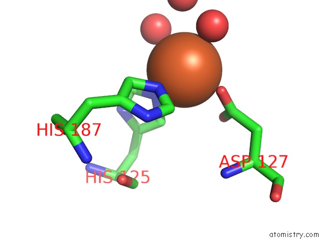
Mono view
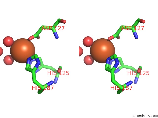
Stereo pair view
Mono view
Stereo pair view
A full contact list of Iron with other atoms in the Fe binding
site number 1 of Crystal Structure of Escherichia Coli Ribosomal Oxygenase Ycfd within 5.0Å range:
|
Reference:
L.M.Van Staalduinen,
S.K.Novakowski,
Z.Jia.
Structure and Functional Analysis of Ycfd, A Novel 2-Oxoglutarate/Fe(2+)-Dependent Oxygenase Involved in Translational Regulation in Escherichia Coli. J.Mol.Biol. V. 426 1898 2014.
ISSN: ISSN 0022-2836
PubMed: 24530688
DOI: 10.1016/J.JMB.2014.02.008
Page generated: Mon Aug 5 08:01:04 2024
ISSN: ISSN 0022-2836
PubMed: 24530688
DOI: 10.1016/J.JMB.2014.02.008
Last articles
Fe in 2YXOFe in 2YRS
Fe in 2YXC
Fe in 2YNM
Fe in 2YVJ
Fe in 2YP1
Fe in 2YU2
Fe in 2YU1
Fe in 2YQB
Fe in 2YOO