Iron »
PDB 4o1w-4oxf »
4o35 »
Iron in PDB 4o35: Crystal Structure of Carbomonoxy Murine Neuroglobin Mutant F106W
Protein crystallography data
The structure of Crystal Structure of Carbomonoxy Murine Neuroglobin Mutant F106W, PDB code: 4o35
was solved by
G.Avella,
C.Savino,
B.Vallone,
with X-Ray Crystallography technique. A brief refinement statistics is given in the table below:
| Resolution Low / High (Å) | 28.18 / 1.80 |
| Space group | H 3 2 |
| Cell size a, b, c (Å), α, β, γ (°) | 89.155, 89.155, 108.622, 90.00, 90.00, 120.00 |
| R / Rfree (%) | 18.8 / 23.4 |
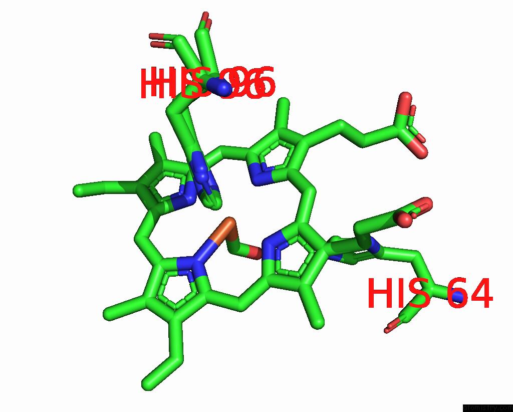
Iron Binding Sites:
The binding sites of Iron atom in the Crystal Structure of Carbomonoxy Murine Neuroglobin Mutant F106W
(pdb code 4o35). This binding sites where shown within
5.0 Angstroms radius around Iron atom.
In total only one binding site of Iron was determined in the Crystal Structure of Carbomonoxy Murine Neuroglobin Mutant F106W, PDB code: 4o35:
In total only one binding site of Iron was determined in the Crystal Structure of Carbomonoxy Murine Neuroglobin Mutant F106W, PDB code: 4o35:
Iron binding site 1 out of 1 in 4o35
Go back to
Iron binding site 1 out
of 1 in the Crystal Structure of Carbomonoxy Murine Neuroglobin Mutant F106W
Mono view

Stereo pair view
Mono view
Stereo pair view
A full contact list of Iron with other atoms in the Fe binding
site number 1 of Crystal Structure of Carbomonoxy Murine Neuroglobin Mutant F106W within 5.0Å range:
|
Reference:
G.Avella,
C.Ardiccioni,
A.Scaglione,
T.Moschetti,
C.Rondinelli,
L.C.Montemiglio,
C.Savino,
A.Giuffre,
M.Brunori,
B.Vallone.
Engineering the Internal Cavity of Neuroglobin Demonstrates the Role of the Haem-Sliding Mechanism. Acta Crystallogr.,Sect.D V. 70 1640 2014.
ISSN: ISSN 0907-4449
PubMed: 24914975
DOI: 10.1107/S1399004714007032
Page generated: Tue Aug 5 13:27:59 2025
ISSN: ISSN 0907-4449
PubMed: 24914975
DOI: 10.1107/S1399004714007032
Last articles
W in 1DV4W in 1FR3
W in 1GUG
W in 1H9R
W in 1H9K
W in 1H0H
W in 1FEZ
W in 1FKA
W in 1E3P
W in 1E18