Iron »
PDB 4o1w-4oxf »
4oo7 »
Iron in PDB 4oo7: The 1.55A Crystal Structure of NAF1 (MINER1): the Redox-Active 2FE-2S Protein
Protein crystallography data
The structure of The 1.55A Crystal Structure of NAF1 (MINER1): the Redox-Active 2FE-2S Protein, PDB code: 4oo7
was solved by
S.Tamir,
Y.Eisenberg-Domovich,
A.R.Colman,
J.T.Stofleth,
C.H.Lipper,
M.L.Paddock,
P.A.Jenning,
O.Livnah,
R.Nechushtai,
with X-Ray Crystallography technique. A brief refinement statistics is given in the table below:
| Resolution Low / High (Å) | 31.38 / 1.65 |
| Space group | P 21 21 21 |
| Cell size a, b, c (Å), α, β, γ (°) | 41.009, 48.652, 73.730, 90.00, 90.00, 90.00 |
| R / Rfree (%) | 13.5 / 15 |
Iron Binding Sites:
The binding sites of Iron atom in the The 1.55A Crystal Structure of NAF1 (MINER1): the Redox-Active 2FE-2S Protein
(pdb code 4oo7). This binding sites where shown within
5.0 Angstroms radius around Iron atom.
In total 4 binding sites of Iron where determined in the The 1.55A Crystal Structure of NAF1 (MINER1): the Redox-Active 2FE-2S Protein, PDB code: 4oo7:
Jump to Iron binding site number: 1; 2; 3; 4;
In total 4 binding sites of Iron where determined in the The 1.55A Crystal Structure of NAF1 (MINER1): the Redox-Active 2FE-2S Protein, PDB code: 4oo7:
Jump to Iron binding site number: 1; 2; 3; 4;
Iron binding site 1 out of 4 in 4oo7
Go back to
Iron binding site 1 out
of 4 in the The 1.55A Crystal Structure of NAF1 (MINER1): the Redox-Active 2FE-2S Protein

Mono view
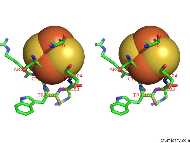
Stereo pair view
Mono view
Stereo pair view
A full contact list of Iron with other atoms in the Fe binding
site number 1 of The 1.55A Crystal Structure of NAF1 (MINER1): the Redox-Active 2FE-2S Protein within 5.0Å range:
|
Iron binding site 2 out of 4 in 4oo7
Go back to
Iron binding site 2 out
of 4 in the The 1.55A Crystal Structure of NAF1 (MINER1): the Redox-Active 2FE-2S Protein
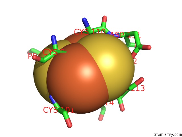
Mono view

Stereo pair view
Mono view
Stereo pair view
A full contact list of Iron with other atoms in the Fe binding
site number 2 of The 1.55A Crystal Structure of NAF1 (MINER1): the Redox-Active 2FE-2S Protein within 5.0Å range:
|
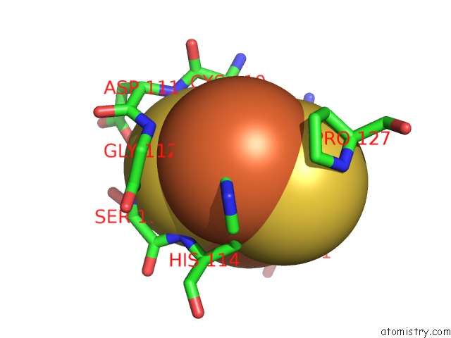
Iron binding site 3 out of 4 in 4oo7
Go back to
Iron binding site 3 out
of 4 in the The 1.55A Crystal Structure of NAF1 (MINER1): the Redox-Active 2FE-2S Protein

Mono view

Stereo pair view
Mono view
Stereo pair view
A full contact list of Iron with other atoms in the Fe binding
site number 3 of The 1.55A Crystal Structure of NAF1 (MINER1): the Redox-Active 2FE-2S Protein within 5.0Å range:
|
Iron binding site 4 out of 4 in 4oo7
Go back to
Iron binding site 4 out
of 4 in the The 1.55A Crystal Structure of NAF1 (MINER1): the Redox-Active 2FE-2S Protein
Mono view
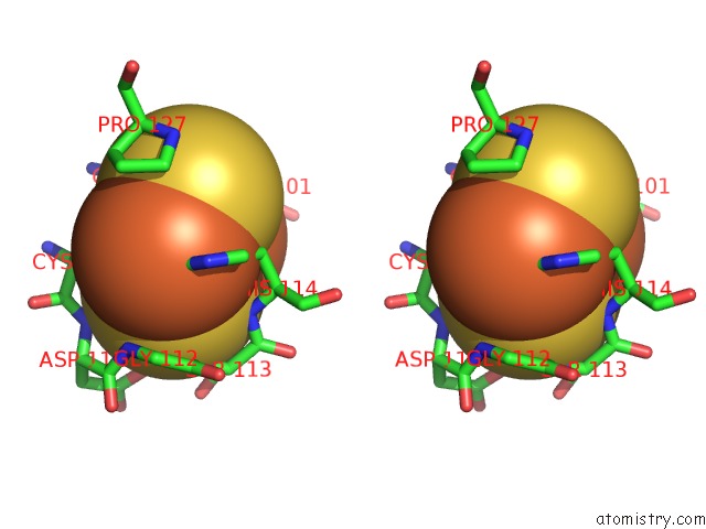
Stereo pair view
Mono view
Stereo pair view
A full contact list of Iron with other atoms in the Fe binding
site number 4 of The 1.55A Crystal Structure of NAF1 (MINER1): the Redox-Active 2FE-2S Protein within 5.0Å range:
|
Reference:
S.Tamir,
Y.Eisenberg-Domovich,
A.R.Conlan,
J.T.Stofleth,
C.H.Lipper,
M.L.Paddock,
R.Mittler,
P.A.Jennings,
O.Livnah,
R.Nechushtai.
A Point Mutation in the [2FE-2S] Cluster Binding Region of the Naf-1 Protein (H114C) Dramatically Hinders the Cluster Donor Properties. Acta Crystallogr.,Sect.D V. 70 1572 2014.
ISSN: ISSN 0907-4449
PubMed: 24914968
DOI: 10.1107/S1399004714005458
Page generated: Tue Aug 5 13:31:04 2025
ISSN: ISSN 0907-4449
PubMed: 24914968
DOI: 10.1107/S1399004714005458
Last articles
W in 1DV4W in 1FR3
W in 1GUG
W in 1H9R
W in 1H9K
W in 1H0H
W in 1FEZ
W in 1FKA
W in 1E3P
W in 1E18