Iron »
PDB 4q4u-4r21 »
4quq »
Iron in PDB 4quq: Crystal Structure of Stachydrine Demethylase in Complex with Azide
Protein crystallography data
The structure of Crystal Structure of Stachydrine Demethylase in Complex with Azide, PDB code: 4quq
was solved by
R.Agarwal,
B.Andi,
A.Gizzi,
J.B.Bonanno,
S.C.Almo,
A.M.Orville,
with X-Ray Crystallography technique. A brief refinement statistics is given in the table below:
| Resolution Low / High (Å) | 33.09 / 2.27 |
| Space group | P 63 2 2 |
| Cell size a, b, c (Å), α, β, γ (°) | 98.440, 98.440, 178.752, 90.00, 90.00, 120.00 |
| R / Rfree (%) | 19.3 / 25.5 |
Other elements in 4quq:
The structure of Crystal Structure of Stachydrine Demethylase in Complex with Azide also contains other interesting chemical elements:
| Cobalt | (Co) | 1 atom |
Iron Binding Sites:
The binding sites of Iron atom in the Crystal Structure of Stachydrine Demethylase in Complex with Azide
(pdb code 4quq). This binding sites where shown within
5.0 Angstroms radius around Iron atom.
In total 3 binding sites of Iron where determined in the Crystal Structure of Stachydrine Demethylase in Complex with Azide, PDB code: 4quq:
Jump to Iron binding site number: 1; 2; 3;
In total 3 binding sites of Iron where determined in the Crystal Structure of Stachydrine Demethylase in Complex with Azide, PDB code: 4quq:
Jump to Iron binding site number: 1; 2; 3;
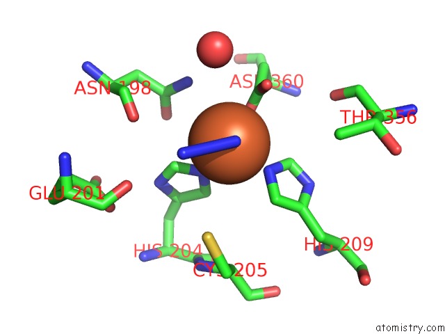
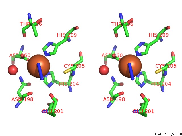
Iron binding site 1 out of 3 in 4quq
Go back to
Iron binding site 1 out
of 3 in the Crystal Structure of Stachydrine Demethylase in Complex with Azide
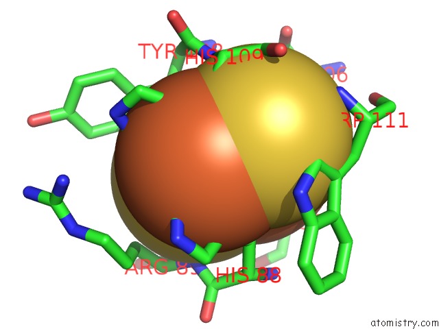
Mono view
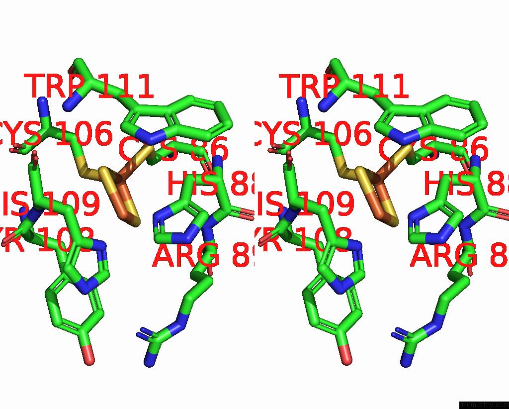
Stereo pair view
Mono view
Stereo pair view
A full contact list of Iron with other atoms in the Fe binding
site number 1 of Crystal Structure of Stachydrine Demethylase in Complex with Azide within 5.0Å range:
|
Iron binding site 2 out of 3 in 4quq
Go back to
Iron binding site 2 out
of 3 in the Crystal Structure of Stachydrine Demethylase in Complex with Azide
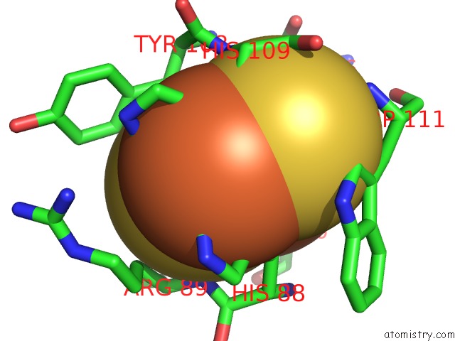
Mono view
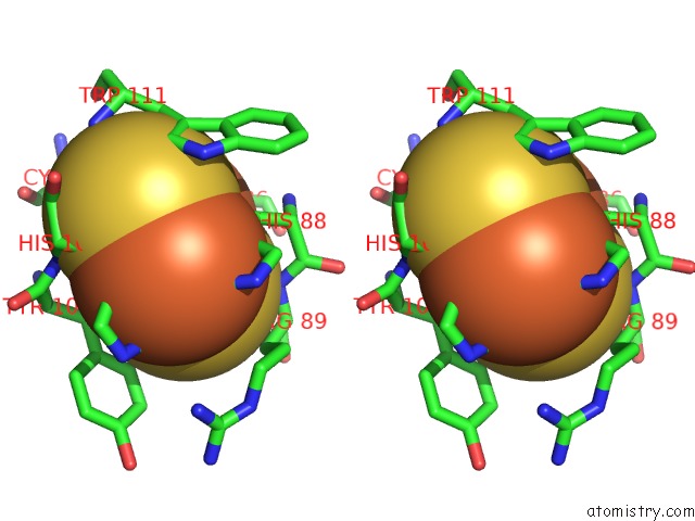
Stereo pair view
Mono view
Stereo pair view
A full contact list of Iron with other atoms in the Fe binding
site number 2 of Crystal Structure of Stachydrine Demethylase in Complex with Azide within 5.0Å range:
|
Iron binding site 3 out of 3 in 4quq
Go back to
Iron binding site 3 out
of 3 in the Crystal Structure of Stachydrine Demethylase in Complex with Azide
Mono view
Stereo pair view
Mono view
Stereo pair view
A full contact list of Iron with other atoms in the Fe binding
site number 3 of Crystal Structure of Stachydrine Demethylase in Complex with Azide within 5.0Å range:
|
Reference:
R.Agarwal,
B.Andi,
A.Gizzi,
J.B.Bonanno,
S.C.Almo,
A.M.Orville.
Tracking Photoelectron Induced in-Crystallo Enzyme Catalysis To Be Published.
Page generated: Tue Aug 5 14:00:54 2025
Last articles
Mg in 5WSAMg in 5WS9
Mg in 5WS8
Mg in 5WS6
Mg in 5WS5
Mg in 5WRT
Mg in 5WRJ
Mg in 5WRI
Mg in 5WQA
Mg in 5WQ0