Iron »
PDB 4r33-4rxn »
4rse »
Iron in PDB 4rse: Crystal Structure of RPE65 in Complex with Mb-001 and Palmitate
Enzymatic activity of Crystal Structure of RPE65 in Complex with Mb-001 and Palmitate
All present enzymatic activity of Crystal Structure of RPE65 in Complex with Mb-001 and Palmitate:
3.1.1.64;
3.1.1.64;
Protein crystallography data
The structure of Crystal Structure of RPE65 in Complex with Mb-001 and Palmitate, PDB code: 4rse
was solved by
P.D.Kiser,
W.Shi,
K.Palczewski,
with X-Ray Crystallography technique. A brief refinement statistics is given in the table below:
| Resolution Low / High (Å) | 47.90 / 2.39 |
| Space group | P 65 |
| Cell size a, b, c (Å), α, β, γ (°) | 175.639, 175.639, 86.611, 90.00, 90.00, 120.00 |
| R / Rfree (%) | 19.6 / 23.6 |
Iron Binding Sites:
The binding sites of Iron atom in the Crystal Structure of RPE65 in Complex with Mb-001 and Palmitate
(pdb code 4rse). This binding sites where shown within
5.0 Angstroms radius around Iron atom.
In total 2 binding sites of Iron where determined in the Crystal Structure of RPE65 in Complex with Mb-001 and Palmitate, PDB code: 4rse:
Jump to Iron binding site number: 1; 2;
In total 2 binding sites of Iron where determined in the Crystal Structure of RPE65 in Complex with Mb-001 and Palmitate, PDB code: 4rse:
Jump to Iron binding site number: 1; 2;
Iron binding site 1 out of 2 in 4rse
Go back to
Iron binding site 1 out
of 2 in the Crystal Structure of RPE65 in Complex with Mb-001 and Palmitate
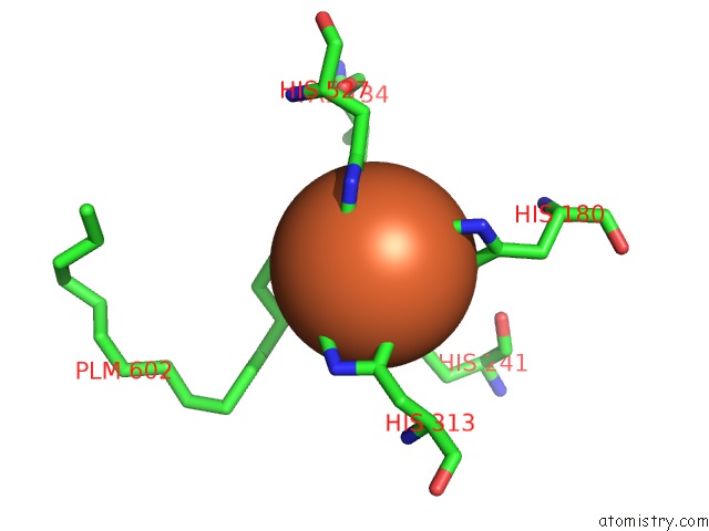
Mono view
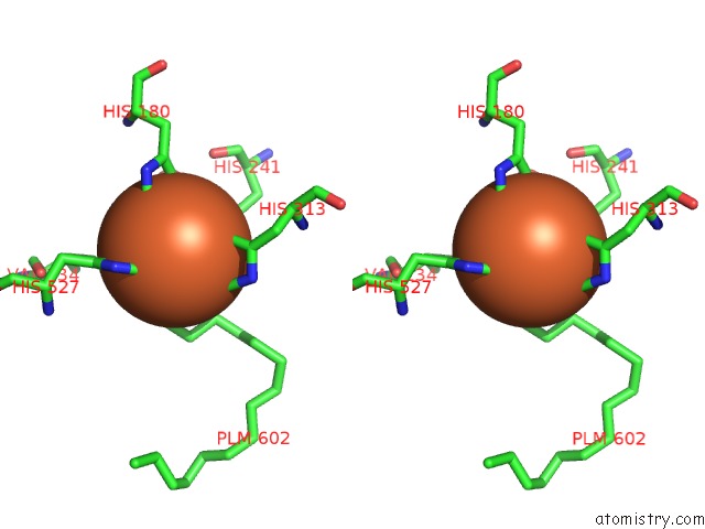
Stereo pair view
Mono view
Stereo pair view
A full contact list of Iron with other atoms in the Fe binding
site number 1 of Crystal Structure of RPE65 in Complex with Mb-001 and Palmitate within 5.0Å range:
|
Iron binding site 2 out of 2 in 4rse
Go back to
Iron binding site 2 out
of 2 in the Crystal Structure of RPE65 in Complex with Mb-001 and Palmitate
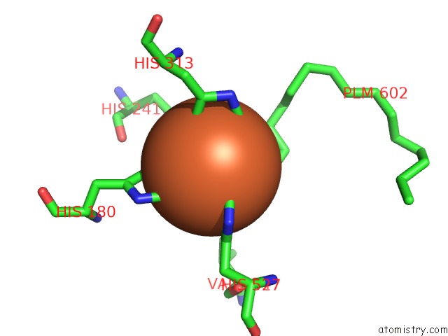
Mono view
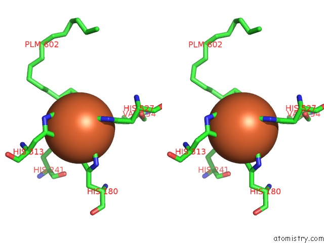
Stereo pair view
Mono view
Stereo pair view
A full contact list of Iron with other atoms in the Fe binding
site number 2 of Crystal Structure of RPE65 in Complex with Mb-001 and Palmitate within 5.0Å range:
|
Reference:
P.D.Kiser,
J.Zhang,
M.Badiee,
Q.Li,
W.Shi,
X.Sui,
M.Golczak,
G.P.Tochtrop,
K.Palczewski.
Catalytic Mechanism of A Retinoid Isomerase Essential For Vertebrate Vision Nat.Chem.Biol. 2015.
ISSN: ESSN 1552-4469
DOI: 10.1038/NCHEMBIO.1799
Page generated: Tue Aug 5 14:18:20 2025
ISSN: ESSN 1552-4469
DOI: 10.1038/NCHEMBIO.1799
Last articles
Mn in 9LJUMn in 9LJW
Mn in 9LJS
Mn in 9LJR
Mn in 9LJT
Mn in 9LJV
Mg in 9UA2
Mg in 9R96
Mg in 9VM1
Mg in 9P01