Iron »
PDB 4ur2-4wgz »
4uzv »
Iron in PDB 4uzv: Structure of A Triple Mutant of Asv-Tftrhb
Protein crystallography data
The structure of Structure of A Triple Mutant of Asv-Tftrhb, PDB code: 4uzv
was solved by
P.Baiocco,
A.Bonamore,
N.Sciamanna,
A.Ilari,
L.Boechi,
A.Boffi,
G.Smulevich,
A.Feis,
with X-Ray Crystallography technique. A brief refinement statistics is given in the table below:
| Resolution Low / High (Å) | 50.00 / 3.40 |
| Space group | P 41 2 2 |
| Cell size a, b, c (Å), α, β, γ (°) | 78.367, 78.367, 89.806, 90.00, 90.00, 90.00 |
| R / Rfree (%) | 29.959 / 34.904 |
Iron Binding Sites:
The binding sites of Iron atom in the Structure of A Triple Mutant of Asv-Tftrhb
(pdb code 4uzv). This binding sites where shown within
5.0 Angstroms radius around Iron atom.
In total only one binding site of Iron was determined in the Structure of A Triple Mutant of Asv-Tftrhb, PDB code: 4uzv:
In total only one binding site of Iron was determined in the Structure of A Triple Mutant of Asv-Tftrhb, PDB code: 4uzv:
Iron binding site 1 out of 1 in 4uzv
Go back to
Iron binding site 1 out
of 1 in the Structure of A Triple Mutant of Asv-Tftrhb
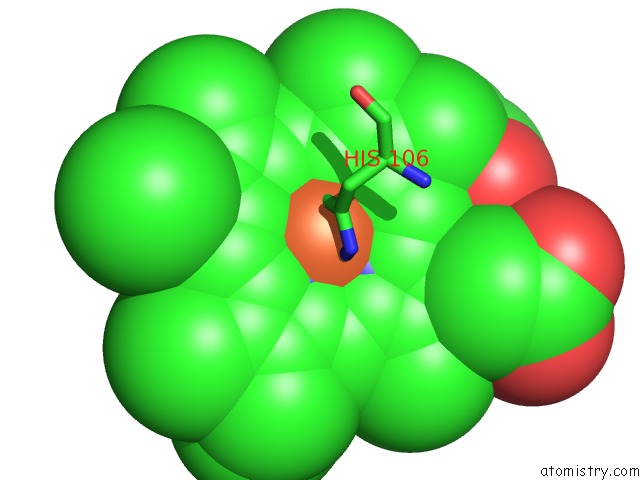
Mono view
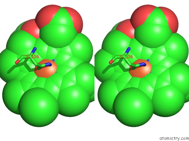
Stereo pair view
Mono view
Stereo pair view
A full contact list of Iron with other atoms in the Fe binding
site number 1 of Structure of A Triple Mutant of Asv-Tftrhb within 5.0Å range:
|
Reference:
B.Patrizi,
A.Lapini,
M.Di Donato,
A.Marcelli,
M.Lima,
R.Righini,
P.Foggi,
P.Baiocco,
A.Bonamore,
A.Boffi.
Role of Local Structure and Dynamics of Small Ligand Migration in Proteins: A Study of A Mutated Truncated Hemoprotein From Thermobifida Fusca By Time Resolved Mir Spectroscopy. J.Phys.Chem.B V. 118 9209 2014.
ISSN: ISSN 1520-6106
PubMed: 25019316
DOI: 10.1021/JP504499B
Page generated: Tue Aug 5 16:26:41 2025
ISSN: ISSN 1520-6106
PubMed: 25019316
DOI: 10.1021/JP504499B
Last articles
Mn in 9LJUMn in 9LJW
Mn in 9LJS
Mn in 9LJR
Mn in 9LJT
Mn in 9LJV
Mg in 9UA2
Mg in 9R96
Mg in 9VM1
Mg in 9P01