Iron »
PDB 4ur2-4wgz »
4w7m »
Iron in PDB 4w7m: Crystal Structure of A Decolorizing Peroxidase (Dyp) From Auricularia Auricula-Judae. W377S Mutant
Enzymatic activity of Crystal Structure of A Decolorizing Peroxidase (Dyp) From Auricularia Auricula-Judae. W377S Mutant
All present enzymatic activity of Crystal Structure of A Decolorizing Peroxidase (Dyp) From Auricularia Auricula-Judae. W377S Mutant:
1.11.1.19;
1.11.1.19;
Protein crystallography data
The structure of Crystal Structure of A Decolorizing Peroxidase (Dyp) From Auricularia Auricula-Judae. W377S Mutant, PDB code: 4w7m
was solved by
F.J.Medrano,
A.Romero,
with X-Ray Crystallography technique. A brief refinement statistics is given in the table below:
| Resolution Low / High (Å) | 41.25 / 1.15 |
| Space group | C 1 2 1 |
| Cell size a, b, c (Å), α, β, γ (°) | 183.960, 56.170, 104.120, 90.00, 118.04, 90.00 |
| R / Rfree (%) | 13.1 / 15 |
Iron Binding Sites:
The binding sites of Iron atom in the Crystal Structure of A Decolorizing Peroxidase (Dyp) From Auricularia Auricula-Judae. W377S Mutant
(pdb code 4w7m). This binding sites where shown within
5.0 Angstroms radius around Iron atom.
In total 2 binding sites of Iron where determined in the Crystal Structure of A Decolorizing Peroxidase (Dyp) From Auricularia Auricula-Judae. W377S Mutant, PDB code: 4w7m:
Jump to Iron binding site number: 1; 2;
In total 2 binding sites of Iron where determined in the Crystal Structure of A Decolorizing Peroxidase (Dyp) From Auricularia Auricula-Judae. W377S Mutant, PDB code: 4w7m:
Jump to Iron binding site number: 1; 2;
Iron binding site 1 out of 2 in 4w7m
Go back to
Iron binding site 1 out
of 2 in the Crystal Structure of A Decolorizing Peroxidase (Dyp) From Auricularia Auricula-Judae. W377S Mutant
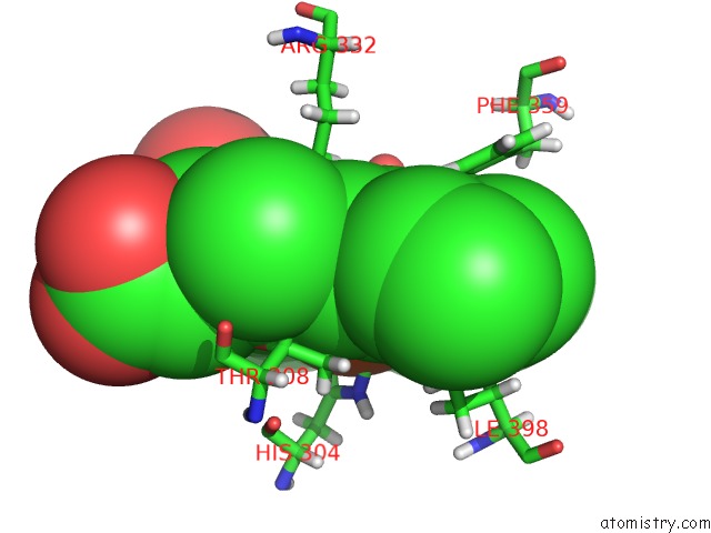
Mono view
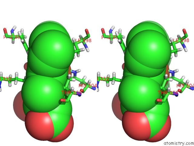
Stereo pair view
Mono view
Stereo pair view
A full contact list of Iron with other atoms in the Fe binding
site number 1 of Crystal Structure of A Decolorizing Peroxidase (Dyp) From Auricularia Auricula-Judae. W377S Mutant within 5.0Å range:
|
Iron binding site 2 out of 2 in 4w7m
Go back to
Iron binding site 2 out
of 2 in the Crystal Structure of A Decolorizing Peroxidase (Dyp) From Auricularia Auricula-Judae. W377S Mutant

Mono view
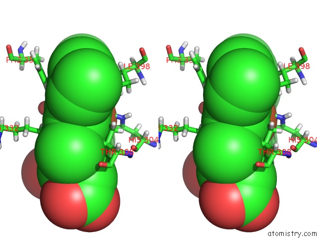
Stereo pair view
Mono view
Stereo pair view
A full contact list of Iron with other atoms in the Fe binding
site number 2 of Crystal Structure of A Decolorizing Peroxidase (Dyp) From Auricularia Auricula-Judae. W377S Mutant within 5.0Å range:
|
Reference:
D.Linde,
R.Pogni,
M.Canellas,
F.Lucas,
V.Guallar,
M.C.Baratto,
A.Sinicropi,
V.Saez-Jimenez,
C.Coscolin,
A.Romero,
F.J.Medrano,
F.J.Ruiz-Duenas,
A.T.Martinez.
Catalytic Surface Radical in Dye-Decolorizing Peroxidase: A Computational, Spectroscopic and Site-Directed Mutagenesis Study. Biochem.J. V. 466 253 2015.
ISSN: ESSN 1470-8728
PubMed: 25495127
DOI: 10.1042/BJ20141211
Page generated: Tue Aug 5 16:35:36 2025
ISSN: ESSN 1470-8728
PubMed: 25495127
DOI: 10.1042/BJ20141211
Last articles
Mn in 9LJUMn in 9LJW
Mn in 9LJS
Mn in 9LJR
Mn in 9LJT
Mn in 9LJV
Mg in 9UA2
Mg in 9R96
Mg in 9VM1
Mg in 9P01