Iron »
PDB 4xry-4yoq »
4y7s »
Iron in PDB 4y7s: Crystal Structure of the Cfem Protein CSA2
Protein crystallography data
The structure of Crystal Structure of the Cfem Protein CSA2, PDB code: 4y7s
was solved by
H.Dvir,
Z.Weissman,
L.Nasser,
D.Hiya,
D.Kornitzer,
with X-Ray Crystallography technique. A brief refinement statistics is given in the table below:
| Resolution Low / High (Å) | 28.67 / 2.00 |
| Space group | P 21 21 21 |
| Cell size a, b, c (Å), α, β, γ (°) | 61.341, 61.208, 97.300, 90.00, 90.00, 90.00 |
| R / Rfree (%) | 15.3 / 19.2 |
Other elements in 4y7s:
The structure of Crystal Structure of the Cfem Protein CSA2 also contains other interesting chemical elements:
| Chlorine | (Cl) | 1 atom |
Iron Binding Sites:
The binding sites of Iron atom in the Crystal Structure of the Cfem Protein CSA2
(pdb code 4y7s). This binding sites where shown within
5.0 Angstroms radius around Iron atom.
In total 3 binding sites of Iron where determined in the Crystal Structure of the Cfem Protein CSA2, PDB code: 4y7s:
Jump to Iron binding site number: 1; 2; 3;
In total 3 binding sites of Iron where determined in the Crystal Structure of the Cfem Protein CSA2, PDB code: 4y7s:
Jump to Iron binding site number: 1; 2; 3;
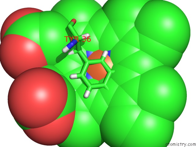
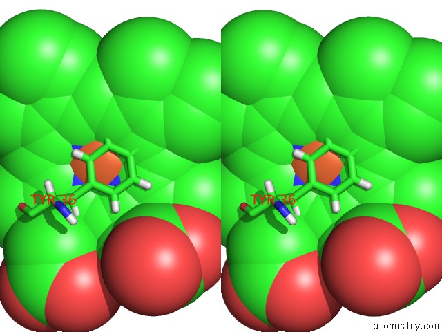
Iron binding site 1 out of 3 in 4y7s
Go back to
Iron binding site 1 out
of 3 in the Crystal Structure of the Cfem Protein CSA2
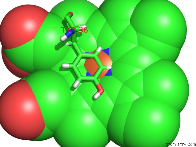
Mono view
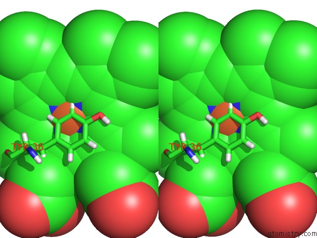
Stereo pair view
Mono view
Stereo pair view
A full contact list of Iron with other atoms in the Fe binding
site number 1 of Crystal Structure of the Cfem Protein CSA2 within 5.0Å range:
|
Iron binding site 2 out of 3 in 4y7s
Go back to
Iron binding site 2 out
of 3 in the Crystal Structure of the Cfem Protein CSA2
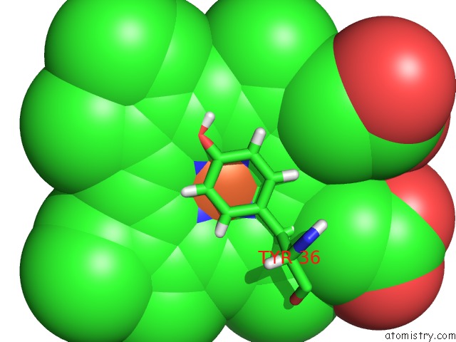
Mono view
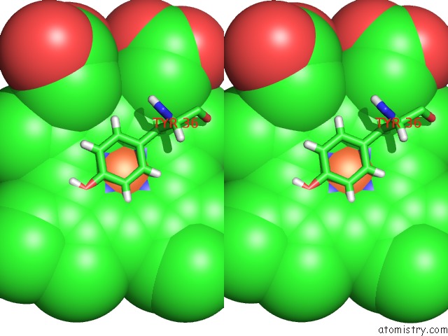
Stereo pair view
Mono view
Stereo pair view
A full contact list of Iron with other atoms in the Fe binding
site number 2 of Crystal Structure of the Cfem Protein CSA2 within 5.0Å range:
|
Iron binding site 3 out of 3 in 4y7s
Go back to
Iron binding site 3 out
of 3 in the Crystal Structure of the Cfem Protein CSA2
Mono view
Stereo pair view
Mono view
Stereo pair view
A full contact list of Iron with other atoms in the Fe binding
site number 3 of Crystal Structure of the Cfem Protein CSA2 within 5.0Å range:
|
Reference:
L.Nasser,
Z.Weissman,
M.Pinsky,
H.Amartely,
H.Dvir,
D.Kornitzer.
Structural Basis of Haem-Iron Acquisition By Fungal Pathogens. Nat Microbiol V. 1 16156 2016.
ISSN: ESSN 2058-5276
PubMed: 27617569
DOI: 10.1038/NMICROBIOL.2016.156
Page generated: Tue Aug 5 17:25:21 2025
ISSN: ESSN 2058-5276
PubMed: 27617569
DOI: 10.1038/NMICROBIOL.2016.156
Last articles
Zn in 9QM9Zn in 9S44
Zn in 9OFE
Zn in 9OFC
Zn in 9OFD
Zn in 9OF1
Zn in 9OFB
Zn in 9N0J
Zn in 9M5X
Zn in 9LGI