Iron »
PDB 4zoh-5ade »
4zoh »
Iron in PDB 4zoh: Crystal Structure of Glyceraldehyde Oxidoreductase
Protein crystallography data
The structure of Crystal Structure of Glyceraldehyde Oxidoreductase, PDB code: 4zoh
was solved by
H.Nishimasu,
S.Fushinobu,
T.Wakagi,
with X-Ray Crystallography technique. A brief refinement statistics is given in the table below:
| Resolution Low / High (Å) | 41.40 / 2.20 |
| Space group | P 65 2 2 |
| Cell size a, b, c (Å), α, β, γ (°) | 143.431, 143.431, 235.509, 90.00, 90.00, 120.00 |
| R / Rfree (%) | 17.7 / 24.2 |
Other elements in 4zoh:
The structure of Crystal Structure of Glyceraldehyde Oxidoreductase also contains other interesting chemical elements:
| Molybdenum | (Mo) | 1 atom |
Iron Binding Sites:
The binding sites of Iron atom in the Crystal Structure of Glyceraldehyde Oxidoreductase
(pdb code 4zoh). This binding sites where shown within
5.0 Angstroms radius around Iron atom.
In total 4 binding sites of Iron where determined in the Crystal Structure of Glyceraldehyde Oxidoreductase, PDB code: 4zoh:
Jump to Iron binding site number: 1; 2; 3; 4;
In total 4 binding sites of Iron where determined in the Crystal Structure of Glyceraldehyde Oxidoreductase, PDB code: 4zoh:
Jump to Iron binding site number: 1; 2; 3; 4;
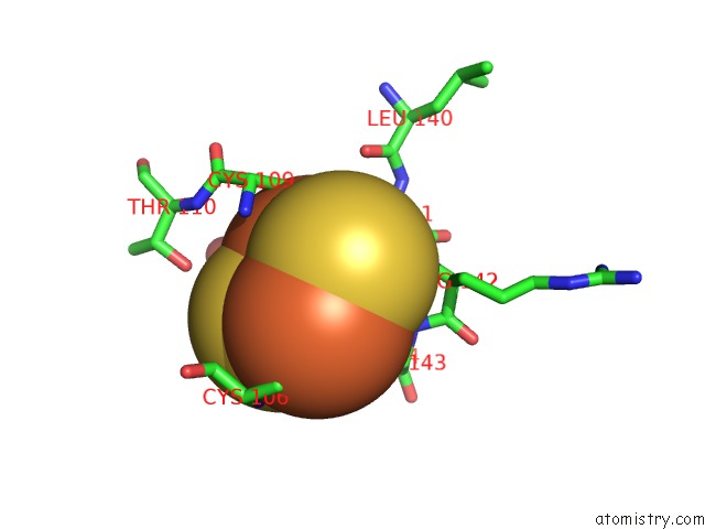
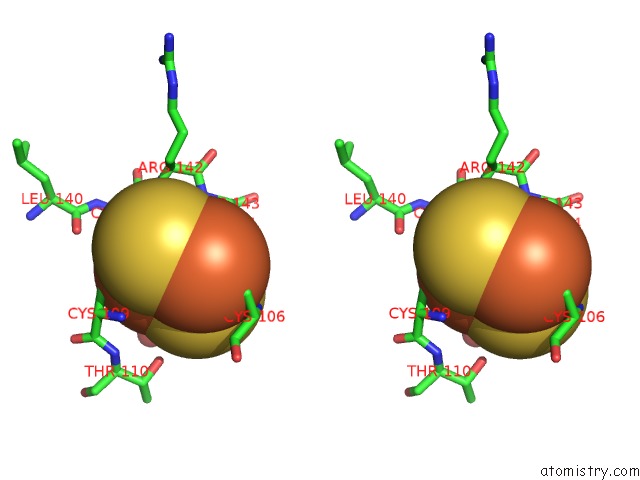
Iron binding site 1 out of 4 in 4zoh
Go back to
Iron binding site 1 out
of 4 in the Crystal Structure of Glyceraldehyde Oxidoreductase
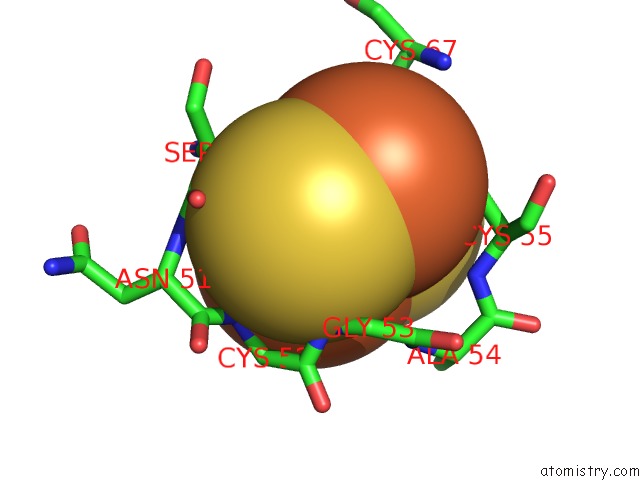
Mono view
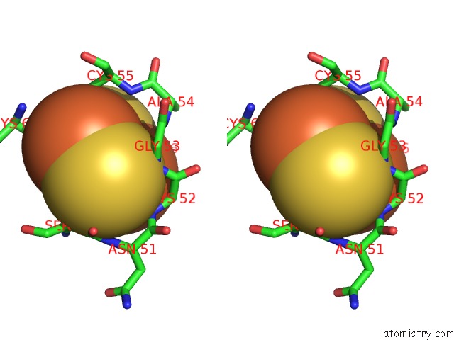
Stereo pair view
Mono view
Stereo pair view
A full contact list of Iron with other atoms in the Fe binding
site number 1 of Crystal Structure of Glyceraldehyde Oxidoreductase within 5.0Å range:
|
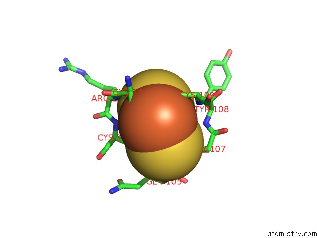
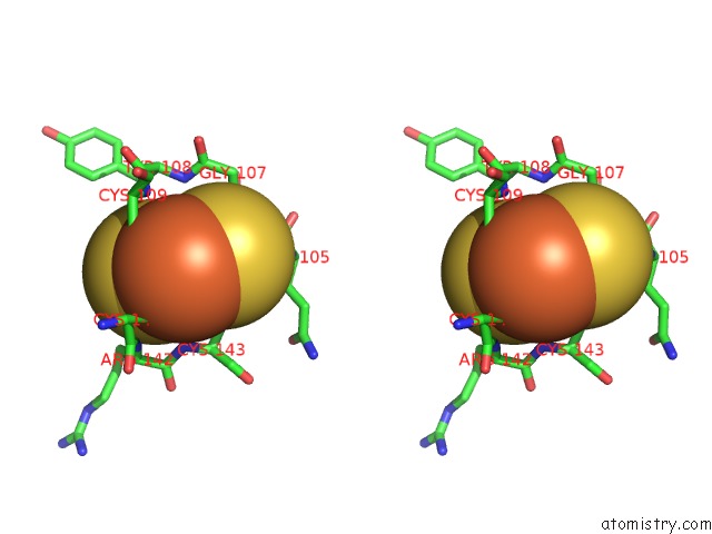
Iron binding site 2 out of 4 in 4zoh
Go back to
Iron binding site 2 out
of 4 in the Crystal Structure of Glyceraldehyde Oxidoreductase
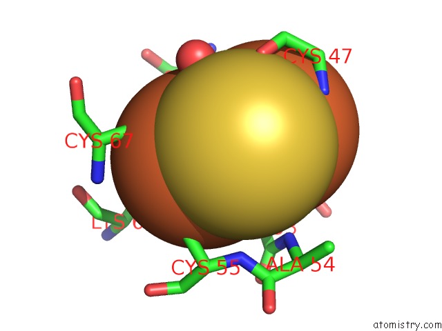
Mono view
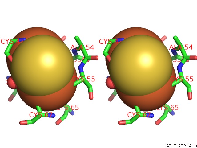
Stereo pair view
Mono view
Stereo pair view
A full contact list of Iron with other atoms in the Fe binding
site number 2 of Crystal Structure of Glyceraldehyde Oxidoreductase within 5.0Å range:
|
Iron binding site 3 out of 4 in 4zoh
Go back to
Iron binding site 3 out
of 4 in the Crystal Structure of Glyceraldehyde Oxidoreductase
Mono view
Stereo pair view
Mono view
Stereo pair view
A full contact list of Iron with other atoms in the Fe binding
site number 3 of Crystal Structure of Glyceraldehyde Oxidoreductase within 5.0Å range:
|
Iron binding site 4 out of 4 in 4zoh
Go back to
Iron binding site 4 out
of 4 in the Crystal Structure of Glyceraldehyde Oxidoreductase
Mono view
Stereo pair view
Mono view
Stereo pair view
A full contact list of Iron with other atoms in the Fe binding
site number 4 of Crystal Structure of Glyceraldehyde Oxidoreductase within 5.0Å range:
|
Reference:
T.Wakagi,
H.Nishimasu,
M.Miyake,
S.Fushinobu.
Archaeal Mo-Containing Glyceraldehyde Oxidoreductase Isozymes Exhibit Diverse Substrate Specificities Through Unique Subunit Assemblies. Plos One V. 11 47333.
ISSN: ESSN 1932-6203
PubMed: 26808202
DOI: 10.1371/JOURNAL.PONE.0147333
Page generated: Tue Aug 5 18:38:37 2025
ISSN: ESSN 1932-6203
PubMed: 26808202
DOI: 10.1371/JOURNAL.PONE.0147333
Last articles
Na in 6HJANa in 6HJX
Na in 6HJ3
Na in 6HIP
Na in 6HHD
Na in 6HE3
Na in 6HE1
Na in 6HEM
Na in 6HDZ
Na in 6HDP