Atomistry » Iron » PDB 5c2i-5cnd » 5c2t
Atomistry »
  Iron »
    PDB 5c2i-5cnd »
      5c2t »

Iron in PDB 5c2t: Crystal Structure of Mitochondrial Rhodoquinol-Fumarate Reductase From Ascaris Suum with Rhodoquinone-2

Enzymatic activity of Crystal Structure of Mitochondrial Rhodoquinol-Fumarate Reductase From Ascaris Suum with Rhodoquinone-2

All present enzymatic activity of Crystal Structure of Mitochondrial Rhodoquinol-Fumarate Reductase From Ascaris Suum with Rhodoquinone-2:
1.3.5.1;

Protein crystallography data

The structure of Crystal Structure of Mitochondrial Rhodoquinol-Fumarate Reductase From Ascaris Suum with Rhodoquinone-2, PDB code: 5c2t was solved by S.Harada, T.Shiba, D.Sato, A.Yamamoto, M.Nagahama, A.Yone, D.K.Inaoka, K.Sakamoto, M.Inoue, T.Honma, K.Kita, with X-Ray Crystallography technique. A brief refinement statistics is given in the table below:

Resolution Low / High (Å) 20.00 / 2.75
Space group P 21 21 21
Cell size a, b, c (Å), α, β, γ (°) 122.813, 123.632, 219.812, 90.00, 90.00, 90.00
R / Rfree (%) 19.6 / 25.2

Iron Binding Sites:

Pages:

>>> Page 1 <<< Page 2, Binding sites: 11 - 20;

Binding sites:

The binding sites of Iron atom in the Crystal Structure of Mitochondrial Rhodoquinol-Fumarate Reductase From Ascaris Suum with Rhodoquinone-2 (pdb code 5c2t). This binding sites where shown within 5.0 Angstroms radius around Iron atom.
In total 20 binding sites of Iron where determined in the Crystal Structure of Mitochondrial Rhodoquinol-Fumarate Reductase From Ascaris Suum with Rhodoquinone-2, PDB code: 5c2t:
Jump to Iron binding site number: 1; 2; 3; 4; 5; 6; 7; 8; 9; 10;

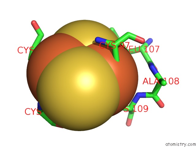
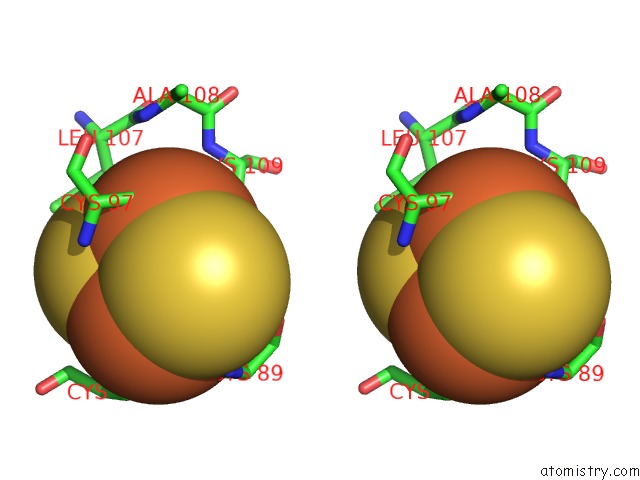
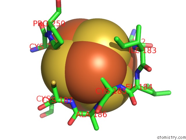
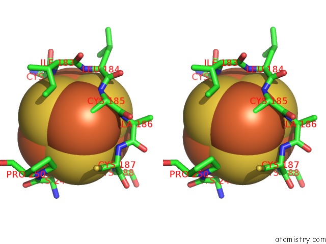
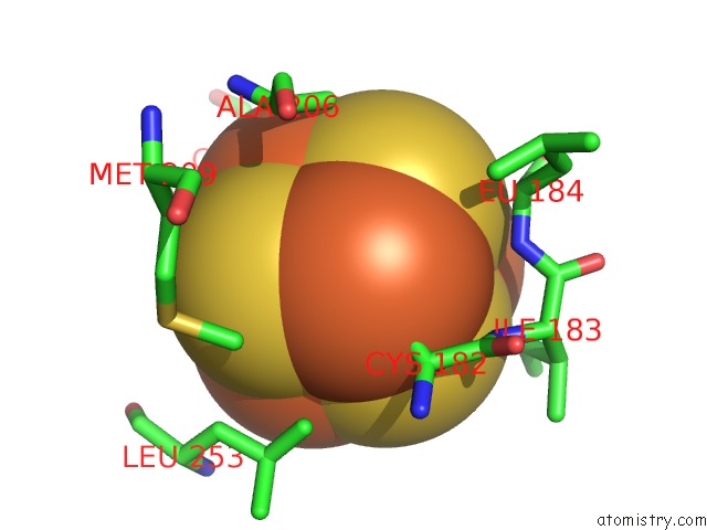
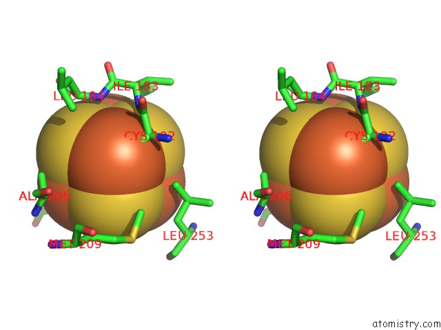
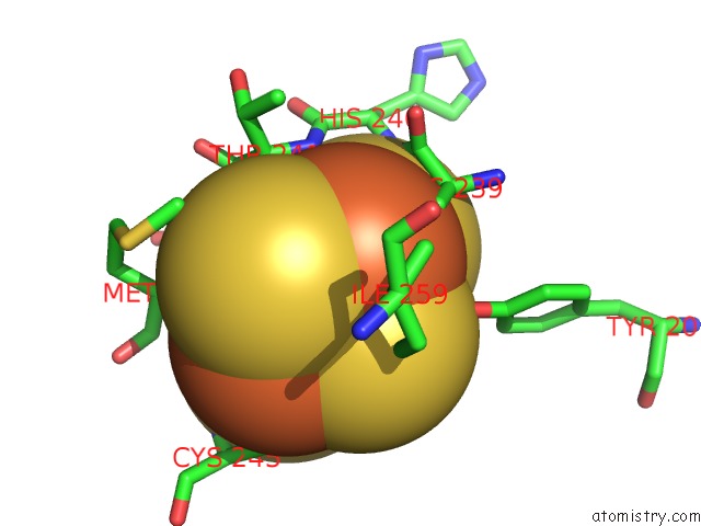
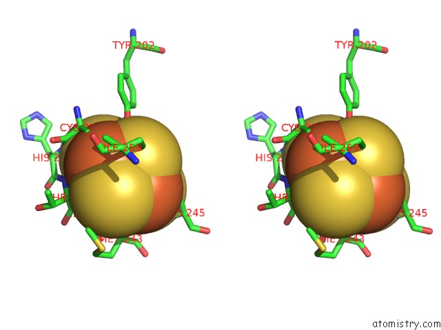
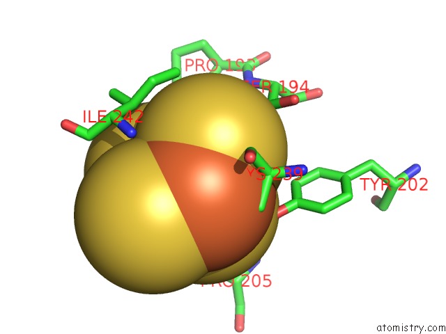
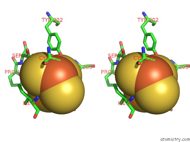
Iron binding site 1 out of 20 in 5c2t

Go back to Iron Binding Sites List in 5c2t
Iron binding site 1 out of 20 in the Crystal Structure of Mitochondrial Rhodoquinol-Fumarate Reductase From Ascaris Suum with Rhodoquinone-2


Mono view


Stereo pair view

A full contact list of Iron with other atoms in the Fe binding site number 1 of Crystal Structure of Mitochondrial Rhodoquinol-Fumarate Reductase From Ascaris Suum with Rhodoquinone-2 within 5.0Å range:
probe atom residue distance (Å) B Occ
B:Fe301

b:24.8
occ:1.00
FE1 B:FES301 0.0 24.8 1.0
SG B:CYS97 2.1 32.9 1.0
S2 B:FES301 2.2 26.8 1.0
S1 B:FES301 2.2 22.8 1.0
SG B:CYS109 2.3 26.1 1.0
FE2 B:FES301 2.6 31.4 1.0
CB B:CYS109 3.0 29.3 1.0
CB B:CYS97 3.3 31.9 1.0
SG B:CYS89 3.8 37.1 1.0
N B:CYS109 4.0 31.4 1.0
N B:CYS97 4.1 32.2 1.0
CA B:CYS109 4.2 30.8 1.0
CA B:CYS97 4.3 31.7 1.0
CB B:LEU107 4.3 31.2 1.0
SG B:CYS94 4.5 29.8 1.0
CA B:GLY92 4.5 30.2 1.0
N B:GLY92 4.7 32.0 1.0
N B:ALA108 4.9 33.5 1.0
CD2 B:LEU107 4.9 30.1 1.0
CG B:LEU107 5.0 30.2 1.0
CD1 B:LEU107 5.0 30.0 1.0

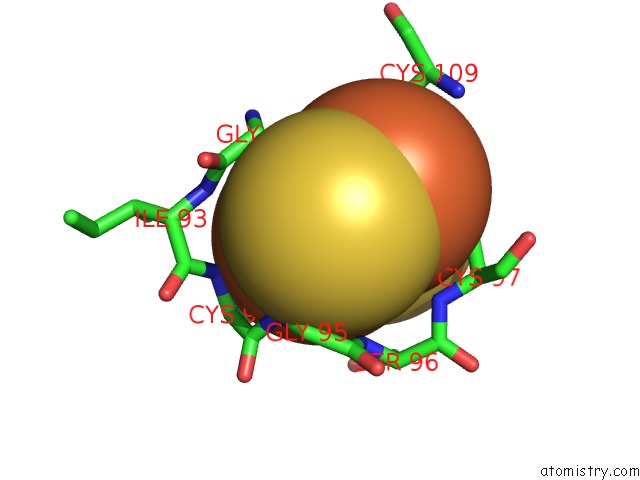
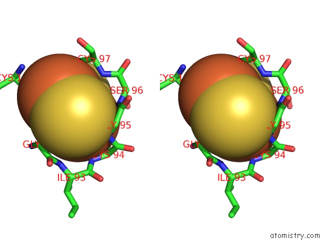
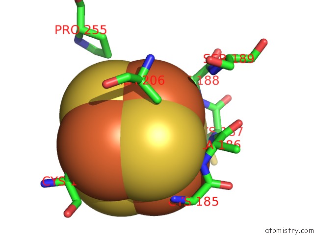
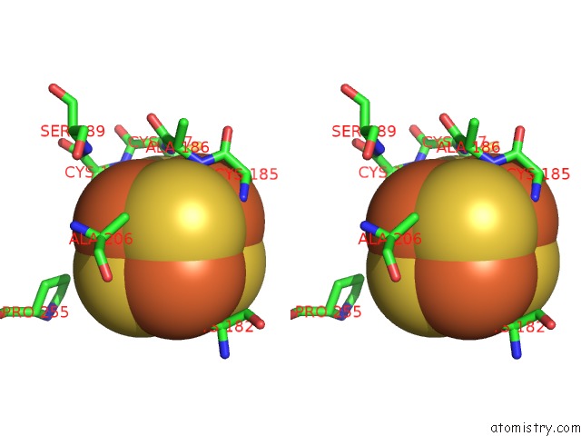
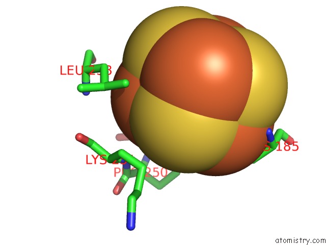
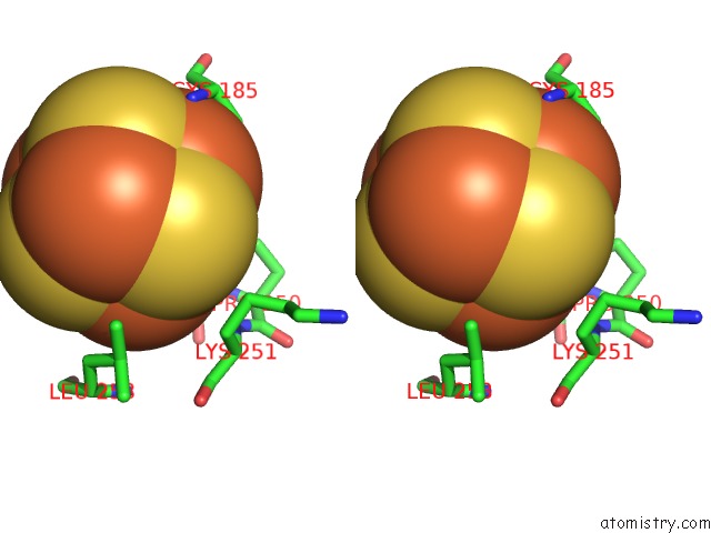
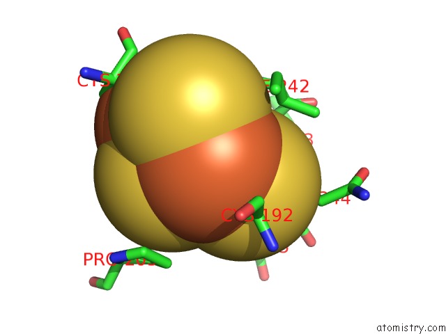
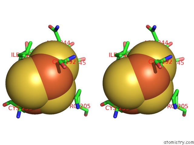
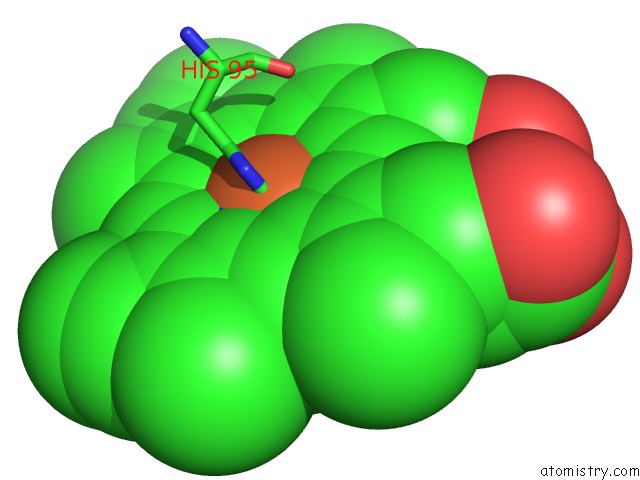
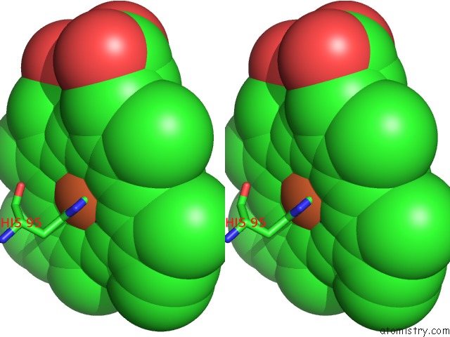
Iron binding site 2 out of 20 in 5c2t

Go back to Iron Binding Sites List in 5c2t
Iron binding site 2 out of 20 in the Crystal Structure of Mitochondrial Rhodoquinol-Fumarate Reductase From Ascaris Suum with Rhodoquinone-2


Mono view


Stereo pair view

A full contact list of Iron with other atoms in the Fe binding site number 2 of Crystal Structure of Mitochondrial Rhodoquinol-Fumarate Reductase From Ascaris Suum with Rhodoquinone-2 within 5.0Å range:
probe atom residue distance (Å) B Occ
B:Fe301

b:31.4
occ:1.00
FE2 B:FES301 0.0 31.4 1.0
SG B:CYS89 2.1 37.1 1.0
S2 B:FES301 2.2 26.8 1.0
SG B:CYS94 2.2 29.8 1.0
S1 B:FES301 2.2 22.8 1.0
FE1 B:FES301 2.6 24.8 1.0
CB B:CYS94 3.4 28.4 1.0
CB B:CYS89 3.5 32.9 1.0
N B:GLY95 3.6 29.3 1.0
N B:CYS94 3.7 26.9 1.0
N B:CYS89 3.8 32.6 1.0
CA B:CYS94 3.9 27.6 1.0
N B:SER96 4.0 31.2 1.0
SG B:CYS109 4.1 26.1 1.0
CA B:CYS89 4.1 34.0 1.0
C B:CYS94 4.2 27.2 1.0
N B:ILE93 4.3 27.7 1.0
SG B:CYS97 4.3 32.9 1.0
CA B:GLY92 4.4 30.2 1.0
C B:CYS89 4.5 36.4 1.0
N B:GLY92 4.5 32.0 1.0
CA B:GLY95 4.5 29.6 1.0
O B:CYS89 4.6 38.4 1.0
OG B:SER96 4.6 39.1 1.0
N B:SER88 4.6 28.8 1.0
N B:CYS97 4.6 32.2 1.0
C B:GLY92 4.7 28.9 1.0
CB B:SER96 4.7 34.5 1.0
C B:GLY95 4.7 30.5 1.0
C B:ILE93 4.7 26.7 1.0
C B:SER88 4.8 31.0 1.0
CA B:SER96 4.9 32.4 1.0
CB B:CYS97 5.0 31.9 1.0

Iron binding site 3 out of 20 in 5c2t

Go back to Iron Binding Sites List in 5c2t
Iron binding site 3 out of 20 in the Crystal Structure of Mitochondrial Rhodoquinol-Fumarate Reductase From Ascaris Suum with Rhodoquinone-2


Mono view


Stereo pair view

A full contact list of Iron with other atoms in the Fe binding site number 3 of Crystal Structure of Mitochondrial Rhodoquinol-Fumarate Reductase From Ascaris Suum with Rhodoquinone-2 within 5.0Å range:
probe atom residue distance (Å) B Occ
B:Fe302

b:30.1
occ:1.00
FE1 B:SF4302 0.0 30.1 1.0
S4 B:SF4302 2.1 26.2 1.0
S3 B:SF4302 2.1 27.1 1.0
S2 B:SF4302 2.1 26.2 1.0
SG B:CYS185 2.1 29.2 1.0
FE2 B:SF4302 3.0 28.3 1.0
FE3 B:SF4302 3.0 28.3 1.0
FE4 B:SF4302 3.1 28.5 1.0
CB B:CYS185 3.4 28.2 1.0
N B:CYS185 3.6 28.3 1.0
N B:ALA186 3.7 29.3 1.0
S1 B:SF4302 3.8 24.4 1.0
CA B:CYS185 3.9 28.2 1.0
N B:CYS187 4.0 29.6 1.0
C B:CYS185 4.2 28.4 1.0
CG1 B:ILE183 4.3 30.3 1.0
CB B:CYS187 4.5 30.6 1.0
CD B:PRO250 4.5 33.1 1.0
CA B:ALA186 4.6 29.3 1.0
SG B:CYS182 4.7 25.6 1.0
C B:LEU184 4.7 28.0 1.0
N B:LEU184 4.7 29.1 1.0
C B:ALA186 4.8 29.5 1.0
CG B:PRO250 4.8 33.7 1.0
CA B:CYS187 4.8 30.1 1.0
N B:CYS188 4.9 30.2 1.0
SG B:CYS249 4.9 25.9 1.0
CA B:LEU184 5.0 28.4 1.0
SG B:CYS188 5.0 31.1 1.0
CD1 B:ILE183 5.0 30.1 1.0

Iron binding site 4 out of 20 in 5c2t

Go back to Iron Binding Sites List in 5c2t
Iron binding site 4 out of 20 in the Crystal Structure of Mitochondrial Rhodoquinol-Fumarate Reductase From Ascaris Suum with Rhodoquinone-2


Mono view


Stereo pair view

A full contact list of Iron with other atoms in the Fe binding site number 4 of Crystal Structure of Mitochondrial Rhodoquinol-Fumarate Reductase From Ascaris Suum with Rhodoquinone-2 within 5.0Å range:
probe atom residue distance (Å) B Occ
B:Fe302

b:28.3
occ:1.00
FE2 B:SF4302 0.0 28.3 1.0
S3 B:SF4302 2.1 27.1 1.0
S4 B:SF4302 2.1 26.2 1.0
S1 B:SF4302 2.1 24.4 1.0
SG B:CYS188 2.3 31.1 1.0
FE3 B:SF4302 2.9 28.3 1.0
FE1 B:SF4302 3.0 30.1 1.0
FE4 B:SF4302 3.0 28.5 1.0
CB B:CYS188 3.1 30.2 1.0
S2 B:SF4302 3.6 26.2 1.0
N B:CYS188 3.8 30.2 1.0
CB B:ALA206 4.0 29.8 1.0
CA B:CYS188 4.1 30.1 1.0
N B:CYS187 4.5 29.6 1.0
CA B:ALA206 4.5 29.6 1.0
SG B:CYS185 4.7 29.2 1.0
SG B:CYS182 4.7 25.6 1.0
N B:ALA186 4.7 29.3 1.0
CA B:ALA186 4.8 29.3 1.0
N B:ALA206 4.9 29.4 1.0
CG B:PRO255 4.9 32.4 1.0
C B:ALA186 4.9 29.5 1.0
C B:CYS187 4.9 29.9 1.0
SG B:CYS249 4.9 25.9 1.0
N B:SER189 5.0 30.2 1.0

Iron binding site 5 out of 20 in 5c2t

Go back to Iron Binding Sites List in 5c2t
Iron binding site 5 out of 20 in the Crystal Structure of Mitochondrial Rhodoquinol-Fumarate Reductase From Ascaris Suum with Rhodoquinone-2


Mono view


Stereo pair view

A full contact list of Iron with other atoms in the Fe binding site number 5 of Crystal Structure of Mitochondrial Rhodoquinol-Fumarate Reductase From Ascaris Suum with Rhodoquinone-2 within 5.0Å range:
probe atom residue distance (Å) B Occ
B:Fe302

b:28.3
occ:1.00
FE3 B:SF4302 0.0 28.3 1.0
S1 B:SF4302 2.1 24.4 1.0
S2 B:SF4302 2.1 26.2 1.0
S4 B:SF4302 2.1 26.2 1.0
SG B:CYS182 2.2 25.6 1.0
FE2 B:SF4302 2.9 28.3 1.0
FE4 B:SF4302 2.9 28.5 1.0
FE1 B:SF4302 3.0 30.1 1.0
CB B:CYS182 3.2 28.5 1.0
S3 B:SF4302 3.6 27.1 1.0
CA B:CYS182 3.7 29.7 1.0
N B:ILE183 4.1 29.4 1.0
C B:CYS182 4.3 29.5 1.0
N B:LEU184 4.4 29.1 1.0
CD1 B:LEU253 4.5 28.7 1.0
CE B:MET209 4.7 40.1 1.0
CB B:ALA206 4.9 29.8 1.0
SG B:CYS188 4.9 31.1 1.0
CA B:LEU184 4.9 28.4 1.0
N B:CYS182 4.9 30.8 1.0

Iron binding site 6 out of 20 in 5c2t

Go back to Iron Binding Sites List in 5c2t
Iron binding site 6 out of 20 in the Crystal Structure of Mitochondrial Rhodoquinol-Fumarate Reductase From Ascaris Suum with Rhodoquinone-2


Mono view


Stereo pair view

A full contact list of Iron with other atoms in the Fe binding site number 6 of Crystal Structure of Mitochondrial Rhodoquinol-Fumarate Reductase From Ascaris Suum with Rhodoquinone-2 within 5.0Å range:
probe atom residue distance (Å) B Occ
B:Fe302

b:28.5
occ:1.00
FE4 B:SF4302 0.0 28.5 1.0
S1 B:SF4302 2.1 24.4 1.0
S3 B:SF4302 2.1 27.1 1.0
S2 B:SF4302 2.1 26.2 1.0
SG B:CYS249 2.2 25.9 1.0
FE3 B:SF4302 2.9 28.3 1.0
FE2 B:SF4302 3.0 28.3 1.0
FE1 B:SF4302 3.1 30.1 1.0
CB B:CYS249 3.2 29.2 1.0
S4 B:SF4302 3.7 26.2 1.0
CA B:CYS249 3.9 29.6 1.0
CD1 B:LEU253 4.2 28.7 1.0
CD B:PRO250 4.3 33.1 1.0
CB B:LEU253 4.5 27.9 1.0
C B:CYS249 4.5 30.5 1.0
CG B:LEU253 4.5 28.0 1.0
N B:PRO250 4.6 32.1 1.0
N B:LYS251 4.7 32.7 1.0
CB B:LYS251 4.8 33.3 1.0
SG B:CYS185 4.9 29.2 1.0

Iron binding site 7 out of 20 in 5c2t

Go back to Iron Binding Sites List in 5c2t
Iron binding site 7 out of 20 in the Crystal Structure of Mitochondrial Rhodoquinol-Fumarate Reductase From Ascaris Suum with Rhodoquinone-2


Mono view


Stereo pair view

A full contact list of Iron with other atoms in the Fe binding site number 7 of Crystal Structure of Mitochondrial Rhodoquinol-Fumarate Reductase From Ascaris Suum with Rhodoquinone-2 within 5.0Å range:
probe atom residue distance (Å) B Occ
B:Fe303

b:37.0
occ:1.00
FE1 B:F3S303 0.0 37.0 1.0
SG B:CYS239 2.1 35.2 1.0
S2 B:F3S303 2.2 36.2 1.0
S3 B:F3S303 2.2 31.7 1.0
S1 B:F3S303 2.2 37.3 1.0
FE3 B:F3S303 2.9 36.1 1.0
FE4 B:F3S303 2.9 35.9 1.0
CB B:CYS239 3.5 35.4 1.0
N B:THR241 3.7 35.9 1.0
CA B:CYS239 3.8 35.9 1.0
OH B:TYR202 3.9 35.2 1.0
N B:HIS240 4.0 36.9 1.0
CA B:THR241 4.0 34.1 1.0
C B:CYS239 4.1 35.9 1.0
S4 B:F3S303 4.1 33.8 1.0
N B:ILE242 4.1 35.8 1.0
CD1 B:ILE259 4.5 34.0 1.0
C B:HIS240 4.6 39.2 1.0
C B:THR241 4.6 35.8 1.0
SG B:CYS245 4.8 27.6 1.0
SG B:CYS192 4.9 29.8 1.0
O B:CYS239 4.9 33.9 1.0
CA B:HIS240 4.9 38.1 1.0
N B:MET243 5.0 36.6 1.0

Iron binding site 8 out of 20 in 5c2t

Go back to Iron Binding Sites List in 5c2t
Iron binding site 8 out of 20 in the Crystal Structure of Mitochondrial Rhodoquinol-Fumarate Reductase From Ascaris Suum with Rhodoquinone-2


Mono view


Stereo pair view

A full contact list of Iron with other atoms in the Fe binding site number 8 of Crystal Structure of Mitochondrial Rhodoquinol-Fumarate Reductase From Ascaris Suum with Rhodoquinone-2 within 5.0Å range:
probe atom residue distance (Å) B Occ
B:Fe303

b:36.1
occ:1.00
FE3 B:F3S303 0.0 36.1 1.0
S3 B:F3S303 2.2 31.7 1.0
S4 B:F3S303 2.2 33.8 1.0
S1 B:F3S303 2.2 37.3 1.0
SG B:CYS245 2.2 27.6 1.0
FE4 B:F3S303 2.8 35.9 1.0
FE1 B:F3S303 2.9 37.0 1.0
CB B:CYS245 3.4 29.9 1.0
N B:CYS245 3.8 32.4 1.0
N B:MET243 3.9 36.6 1.0
S2 B:F3S303 3.9 36.2 1.0
CA B:MET243 4.2 38.4 1.0
CA B:CYS245 4.2 30.9 1.0
N B:ASN244 4.3 37.5 1.0
C B:MET243 4.5 38.0 1.0
SG B:CYS239 4.7 35.2 1.0
N B:ILE242 4.7 35.8 1.0
CB B:ALA256 4.8 33.9 1.0
CB B:PRO205 4.9 29.5 1.0
CG B:PRO205 4.9 29.2 1.0
C B:ILE242 4.9 35.4 1.0
SG B:CYS192 4.9 29.8 1.0
C B:ASN244 4.9 33.9 1.0
CB B:CYS192 5.0 31.2 1.0

Iron binding site 9 out of 20 in 5c2t

Go back to Iron Binding Sites List in 5c2t
Iron binding site 9 out of 20 in the Crystal Structure of Mitochondrial Rhodoquinol-Fumarate Reductase From Ascaris Suum with Rhodoquinone-2


Mono view


Stereo pair view

A full contact list of Iron with other atoms in the Fe binding site number 9 of Crystal Structure of Mitochondrial Rhodoquinol-Fumarate Reductase From Ascaris Suum with Rhodoquinone-2 within 5.0Å range:
probe atom residue distance (Å) B Occ
B:Fe303

b:35.9
occ:1.00
FE4 B:F3S303 0.0 35.9 1.0
S4 B:F3S303 2.2 33.8 1.0
S3 B:F3S303 2.2 31.7 1.0
S2 B:F3S303 2.2 36.2 1.0
SG B:CYS192 2.4 29.8 1.0
FE3 B:F3S303 2.8 36.1 1.0
FE1 B:F3S303 2.9 37.0 1.0
CB B:CYS192 3.1 31.2 1.0
CA B:CYS192 3.8 32.0 1.0
S1 B:F3S303 4.0 37.3 1.0
CB B:ILE242 4.4 32.6 1.0
OH B:TYR202 4.6 35.2 1.0
C B:CYS192 4.6 34.1 1.0
CD1 B:ILE242 4.6 32.4 1.0
CB B:SER194 4.7 34.5 1.0
SG B:CYS245 4.7 27.6 1.0
N B:ILE242 4.8 35.8 1.0
CG1 B:ILE242 4.8 33.2 1.0
SG B:CYS239 4.9 35.2 1.0
CD B:PRO205 4.9 29.5 1.0
CE2 B:TYR202 4.9 33.3 1.0
N B:PRO193 5.0 35.3 1.0

Iron binding site 10 out of 20 in 5c2t

Go back to Iron Binding Sites List in 5c2t
Iron binding site 10 out of 20 in the Crystal Structure of Mitochondrial Rhodoquinol-Fumarate Reductase From Ascaris Suum with Rhodoquinone-2


Mono view


Stereo pair view

A full contact list of Iron with other atoms in the Fe binding site number 10 of Crystal Structure of Mitochondrial Rhodoquinol-Fumarate Reductase From Ascaris Suum with Rhodoquinone-2 within 5.0Å range:
probe atom residue distance (Å) B Occ
C:Fe201

b:49.2
occ:1.00
FE C:HEM201 0.0 49.2 1.0
ND C:HEM201 1.9 52.4 1.0
NA C:HEM201 2.0 54.6 1.0
NC C:HEM201 2.1 53.2 1.0
NB C:HEM201 2.1 55.5 1.0
NE2 D:HIS95 2.1 38.8 1.0
NE2 C:HIS131 2.2 39.0 1.0
C4D C:HEM201 2.9 53.4 1.0
C1D C:HEM201 2.9 51.8 1.0
C1A C:HEM201 3.0 52.6 1.0
C4A C:HEM201 3.0 55.6 1.0
C1C C:HEM201 3.1 53.8 1.0
C1B C:HEM201 3.1 57.0 1.0
C4B C:HEM201 3.1 55.4 1.0
C4C C:HEM201 3.1 53.6 1.0
CD2 C:HIS131 3.1 39.7 1.0
CE1 D:HIS95 3.1 40.6 1.0
CD2 D:HIS95 3.1 41.7 1.0
CE1 C:HIS131 3.2 40.4 1.0
CHA C:HEM201 3.4 53.6 1.0
CHD C:HEM201 3.4 53.9 1.0
CHB C:HEM201 3.4 56.7 1.0
CHC C:HEM201 3.4 53.7 1.0
C3D C:HEM201 4.2 50.5 1.0
C2D C:HEM201 4.2 51.0 1.0
ND1 D:HIS95 4.2 41.2 1.0
C3A C:HEM201 4.2 54.3 1.0
C2A C:HEM201 4.2 51.3 1.0
CG D:HIS95 4.2 42.8 1.0
C2C C:HEM201 4.2 53.3 1.0
ND1 C:HIS131 4.3 40.0 1.0
CG C:HIS131 4.3 41.4 1.0
C3C C:HEM201 4.3 53.7 1.0
C2B C:HEM201 4.3 59.4 1.0
C3B C:HEM201 4.3 61.6 1.0
NE2 C:HIS75 4.7 46.3 1.0
CE1 C:HIS75 4.9 45.3 1.0

Reference:

D.K.Inaoka, T.Shiba, D.Sato, E.O.Balogun, T.Sasaki, M.Nagahama, M.Oda, S.Matsuoka, J.Ohmori, T.Honma, M.Inoue, K.Kita, S.Harada. Structural Insights Into the Molecular Design of Flutolanil Derivatives Targeted For Fumarate Respiration of Parasite Mitochondria Int J Mol Sci V. 16 15287 2015.
ISSN: ESSN 1422-0067
PubMed: 26198225
DOI: 10.3390/IJMS160715287
Page generated: Tue Aug 5 19:38:56 2025

Last articles

Na in 6TBI
Na in 6TCJ
Na in 6TBT
Na in 6TBN
Na in 6TBF
Na in 6TBH
Na in 6TBG
Na in 6TB7
Na in 6TB1
Na in 6T99
© Copyright 2008-2020 by atomistry.com
Home   |    Site Map   |    Copyright   |    Contact us   |    Privacy