Iron »
PDB 5g6f-5h8y »
5gj3 »
Iron in PDB 5gj3: Periplasmic Heme-Binding Protein Rhut From Roseiflexus Sp. Rs-1 in Two-Heme Bound Form (Holo-2)
Protein crystallography data
The structure of Periplasmic Heme-Binding Protein Rhut From Roseiflexus Sp. Rs-1 in Two-Heme Bound Form (Holo-2), PDB code: 5gj3
was solved by
M.M.Rahman,
Y.Naoe,
N.Nakamura,
Y.Shiro,
H.Sugimoto,
with X-Ray Crystallography technique. A brief refinement statistics is given in the table below:
| Resolution Low / High (Å) | 43.21 / 2.00 |
| Space group | C 2 2 21 |
| Cell size a, b, c (Å), α, β, γ (°) | 67.326, 86.338, 118.806, 90.00, 90.00, 90.00 |
| R / Rfree (%) | 18.7 / 22.4 |
Other elements in 5gj3:
The structure of Periplasmic Heme-Binding Protein Rhut From Roseiflexus Sp. Rs-1 in Two-Heme Bound Form (Holo-2) also contains other interesting chemical elements:
| Zinc | (Zn) | 4 atoms |
Iron Binding Sites:
The binding sites of Iron atom in the Periplasmic Heme-Binding Protein Rhut From Roseiflexus Sp. Rs-1 in Two-Heme Bound Form (Holo-2)
(pdb code 5gj3). This binding sites where shown within
5.0 Angstroms radius around Iron atom.
In total 2 binding sites of Iron where determined in the Periplasmic Heme-Binding Protein Rhut From Roseiflexus Sp. Rs-1 in Two-Heme Bound Form (Holo-2), PDB code: 5gj3:
Jump to Iron binding site number: 1; 2;
In total 2 binding sites of Iron where determined in the Periplasmic Heme-Binding Protein Rhut From Roseiflexus Sp. Rs-1 in Two-Heme Bound Form (Holo-2), PDB code: 5gj3:
Jump to Iron binding site number: 1; 2;
Iron binding site 1 out of 2 in 5gj3
Go back to
Iron binding site 1 out
of 2 in the Periplasmic Heme-Binding Protein Rhut From Roseiflexus Sp. Rs-1 in Two-Heme Bound Form (Holo-2)
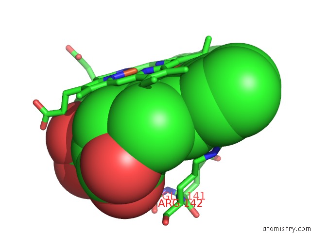
Mono view
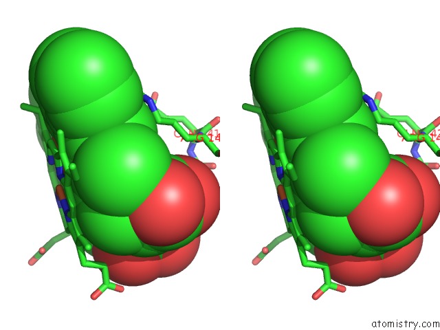
Stereo pair view
Mono view
Stereo pair view
A full contact list of Iron with other atoms in the Fe binding
site number 1 of Periplasmic Heme-Binding Protein Rhut From Roseiflexus Sp. Rs-1 in Two-Heme Bound Form (Holo-2) within 5.0Å range:
|
Iron binding site 2 out of 2 in 5gj3
Go back to
Iron binding site 2 out
of 2 in the Periplasmic Heme-Binding Protein Rhut From Roseiflexus Sp. Rs-1 in Two-Heme Bound Form (Holo-2)
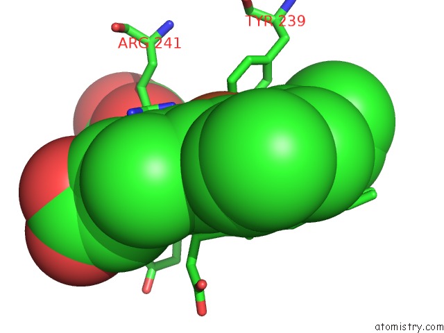
Mono view
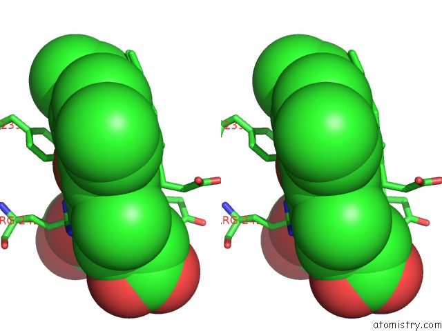
Stereo pair view
Mono view
Stereo pair view
A full contact list of Iron with other atoms in the Fe binding
site number 2 of Periplasmic Heme-Binding Protein Rhut From Roseiflexus Sp. Rs-1 in Two-Heme Bound Form (Holo-2) within 5.0Å range:
|
Reference:
Y.Naoe,
N.Nakamura,
M.M.Rahman,
T.Tosha,
S.Nagatoishi,
K.Tsumoto,
Y.Shiro,
H.Sugimoto.
Structural Basis For Binding and Transfer of Heme in Bacterial Heme-Acquisition Systems. Proteins V. 85 2217 2017.
ISSN: ESSN 1097-0134
PubMed: 28913898
DOI: 10.1002/PROT.25386
Page generated: Tue Aug 5 21:40:50 2025
ISSN: ESSN 1097-0134
PubMed: 28913898
DOI: 10.1002/PROT.25386
Last articles
Mg in 9GURMg in 9GRE
Mg in 9GTK
Mg in 9GU5
Mg in 9GOB
Mg in 9GO5
Mg in 9GMZ
Mg in 9GQO
Mg in 9GMX
Mg in 9GMW