Iron »
PDB 5g6f-5h8y »
5gxg »
Iron in PDB 5gxg: High-Resolution Crystal Structure of the Electron Transfer Complex of Cytochrome P450CAM with Putidaredoxin
Enzymatic activity of High-Resolution Crystal Structure of the Electron Transfer Complex of Cytochrome P450CAM with Putidaredoxin
All present enzymatic activity of High-Resolution Crystal Structure of the Electron Transfer Complex of Cytochrome P450CAM with Putidaredoxin:
1.14.15.1;
1.14.15.1;
Protein crystallography data
The structure of High-Resolution Crystal Structure of the Electron Transfer Complex of Cytochrome P450CAM with Putidaredoxin, PDB code: 5gxg
was solved by
Y.Kikui,
Y.Hiruma,
M.Ubbink,
M.Nojiri,
with X-Ray Crystallography technique. A brief refinement statistics is given in the table below:
| Resolution Low / High (Å) | 59.66 / 1.70 |
| Space group | C 1 2 1 |
| Cell size a, b, c (Å), α, β, γ (°) | 101.490, 77.956, 59.968, 90.00, 95.51, 90.00 |
| R / Rfree (%) | 15.6 / 19.2 |
Iron Binding Sites:
The binding sites of Iron atom in the High-Resolution Crystal Structure of the Electron Transfer Complex of Cytochrome P450CAM with Putidaredoxin
(pdb code 5gxg). This binding sites where shown within
5.0 Angstroms radius around Iron atom.
In total 3 binding sites of Iron where determined in the High-Resolution Crystal Structure of the Electron Transfer Complex of Cytochrome P450CAM with Putidaredoxin, PDB code: 5gxg:
Jump to Iron binding site number: 1; 2; 3;
In total 3 binding sites of Iron where determined in the High-Resolution Crystal Structure of the Electron Transfer Complex of Cytochrome P450CAM with Putidaredoxin, PDB code: 5gxg:
Jump to Iron binding site number: 1; 2; 3;
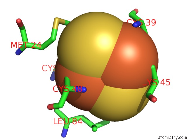
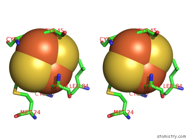
Iron binding site 1 out of 3 in 5gxg
Go back to
Iron binding site 1 out
of 3 in the High-Resolution Crystal Structure of the Electron Transfer Complex of Cytochrome P450CAM with Putidaredoxin
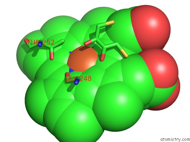
Mono view
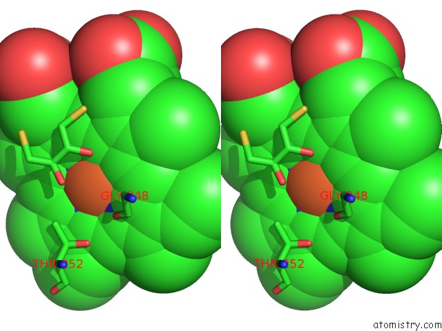
Stereo pair view
Mono view
Stereo pair view
A full contact list of Iron with other atoms in the Fe binding
site number 1 of High-Resolution Crystal Structure of the Electron Transfer Complex of Cytochrome P450CAM with Putidaredoxin within 5.0Å range:
|
Iron binding site 2 out of 3 in 5gxg
Go back to
Iron binding site 2 out
of 3 in the High-Resolution Crystal Structure of the Electron Transfer Complex of Cytochrome P450CAM with Putidaredoxin
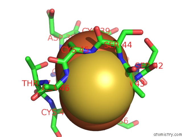
Mono view
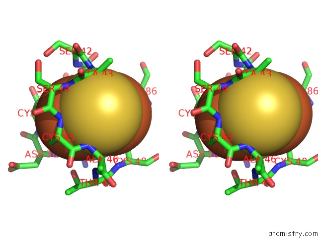
Stereo pair view
Mono view
Stereo pair view
A full contact list of Iron with other atoms in the Fe binding
site number 2 of High-Resolution Crystal Structure of the Electron Transfer Complex of Cytochrome P450CAM with Putidaredoxin within 5.0Å range:
|
Iron binding site 3 out of 3 in 5gxg
Go back to
Iron binding site 3 out
of 3 in the High-Resolution Crystal Structure of the Electron Transfer Complex of Cytochrome P450CAM with Putidaredoxin
Mono view
Stereo pair view
Mono view
Stereo pair view
A full contact list of Iron with other atoms in the Fe binding
site number 3 of High-Resolution Crystal Structure of the Electron Transfer Complex of Cytochrome P450CAM with Putidaredoxin within 5.0Å range:
|
Reference:
W.Andraojc,
Y.Hiruma,
W.M.Liu,
E.Ravera,
M.Nojiri,
G.Parigi,
C.Luchinat,
M.Ubbink.
Identification of Productive and Futile Encounters in An Electron Transfer Protein Complex Proc. Natl. Acad. Sci. V. 114 E1840 2017U.S.A..
ISSN: ESSN 1091-6490
PubMed: 28223532
DOI: 10.1073/PNAS.1616813114
Page generated: Tue Aug 5 21:45:06 2025
ISSN: ESSN 1091-6490
PubMed: 28223532
DOI: 10.1073/PNAS.1616813114
Last articles
K in 9NESK in 9PHG
K in 9NEI
K in 9NED
K in 9NEC
K in 9NEG
K in 9CWU
K in 9CVB
K in 9CVA
K in 9COM