Iron »
PDB 5g6f-5h8y »
5h8c »
Iron in PDB 5h8c: Truncated Xpd
Protein crystallography data
The structure of Truncated Xpd, PDB code: 5h8c
was solved by
J.H.Naismith,
D.Constantinescu,
with X-Ray Crystallography technique. A brief refinement statistics is given in the table below:
| Resolution Low / High (Å) | 29.00 / 2.29 |
| Space group | P 21 21 21 |
| Cell size a, b, c (Å), α, β, γ (°) | 64.800, 77.800, 99.540, 90.00, 90.00, 90.00 |
| R / Rfree (%) | 20.3 / 23.9 |
Iron Binding Sites:
The binding sites of Iron atom in the Truncated Xpd
(pdb code 5h8c). This binding sites where shown within
5.0 Angstroms radius around Iron atom.
In total 4 binding sites of Iron where determined in the Truncated Xpd, PDB code: 5h8c:
Jump to Iron binding site number: 1; 2; 3; 4;
In total 4 binding sites of Iron where determined in the Truncated Xpd, PDB code: 5h8c:
Jump to Iron binding site number: 1; 2; 3; 4;
Iron binding site 1 out of 4 in 5h8c
Go back to
Iron binding site 1 out
of 4 in the Truncated Xpd
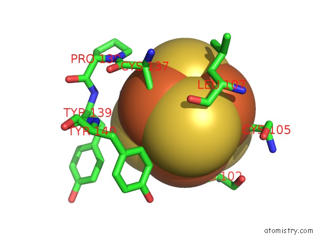
Mono view
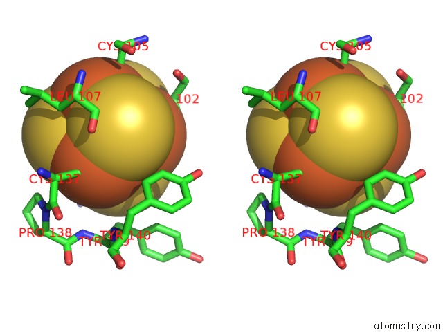
Stereo pair view
Mono view
Stereo pair view
A full contact list of Iron with other atoms in the Fe binding
site number 1 of Truncated Xpd within 5.0Å range:
|
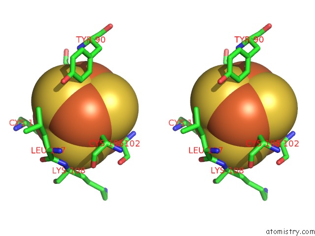
Iron binding site 2 out of 4 in 5h8c
Go back to
Iron binding site 2 out
of 4 in the Truncated Xpd

Mono view

Stereo pair view
Mono view
Stereo pair view
A full contact list of Iron with other atoms in the Fe binding
site number 2 of Truncated Xpd within 5.0Å range:
|
Iron binding site 3 out of 4 in 5h8c
Go back to
Iron binding site 3 out
of 4 in the Truncated Xpd

Mono view

Stereo pair view
Mono view
Stereo pair view
A full contact list of Iron with other atoms in the Fe binding
site number 3 of Truncated Xpd within 5.0Å range:
|
Iron binding site 4 out of 4 in 5h8c
Go back to
Iron binding site 4 out
of 4 in the Truncated Xpd

Mono view
Stereo pair view
Mono view
Stereo pair view
A full contact list of Iron with other atoms in the Fe binding
site number 4 of Truncated Xpd within 5.0Å range:
|
Reference:
D.Constantinescu-Aruxandei,
B.Petrovic-Stojanovska,
J.C.Penedo,
M.F.White,
J.H.Naismith.
Mechanism of Dna Loading By the Dna Repair Helicase Xpd. Nucleic Acids Res. V. 44 2806 2016.
ISSN: ESSN 1362-4962
PubMed: 26896802
DOI: 10.1093/NAR/GKW102
Page generated: Tue Aug 5 21:47:26 2025
ISSN: ESSN 1362-4962
PubMed: 26896802
DOI: 10.1093/NAR/GKW102
Last articles
K in 9NESK in 9PHG
K in 9NEI
K in 9NED
K in 9NEC
K in 9NEG
K in 9CWU
K in 9CVB
K in 9CVA
K in 9COM