Iron »
PDB 5h92-5ibe »
5hr7 »
Iron in PDB 5hr7: X-Ray Crystal Structure of C118A Rlmn From Escherichia Coli with Cross-Linked in Vitro Transcribed Trna
Enzymatic activity of X-Ray Crystal Structure of C118A Rlmn From Escherichia Coli with Cross-Linked in Vitro Transcribed Trna
All present enzymatic activity of X-Ray Crystal Structure of C118A Rlmn From Escherichia Coli with Cross-Linked in Vitro Transcribed Trna:
2.1.1.192;
2.1.1.192;
Protein crystallography data
The structure of X-Ray Crystal Structure of C118A Rlmn From Escherichia Coli with Cross-Linked in Vitro Transcribed Trna, PDB code: 5hr7
was solved by
E.L.Schwalm,
T.L.Grove,
S.J.Booker,
A.K.Boal,
with X-Ray Crystallography technique. A brief refinement statistics is given in the table below:
| Resolution Low / High (Å) | 50.00 / 2.40 |
| Space group | P 1 21 1 |
| Cell size a, b, c (Å), α, β, γ (°) | 90.717, 70.383, 151.810, 90.00, 90.11, 90.00 |
| R / Rfree (%) | 21.6 / 24.8 |
Other elements in 5hr7:
The structure of X-Ray Crystal Structure of C118A Rlmn From Escherichia Coli with Cross-Linked in Vitro Transcribed Trna also contains other interesting chemical elements:
| Magnesium | (Mg) | 7 atoms |
| Arsenic | (As) | 2 atoms |
Iron Binding Sites:
The binding sites of Iron atom in the X-Ray Crystal Structure of C118A Rlmn From Escherichia Coli with Cross-Linked in Vitro Transcribed Trna
(pdb code 5hr7). This binding sites where shown within
5.0 Angstroms radius around Iron atom.
In total 8 binding sites of Iron where determined in the X-Ray Crystal Structure of C118A Rlmn From Escherichia Coli with Cross-Linked in Vitro Transcribed Trna, PDB code: 5hr7:
Jump to Iron binding site number: 1; 2; 3; 4; 5; 6; 7; 8;
In total 8 binding sites of Iron where determined in the X-Ray Crystal Structure of C118A Rlmn From Escherichia Coli with Cross-Linked in Vitro Transcribed Trna, PDB code: 5hr7:
Jump to Iron binding site number: 1; 2; 3; 4; 5; 6; 7; 8;
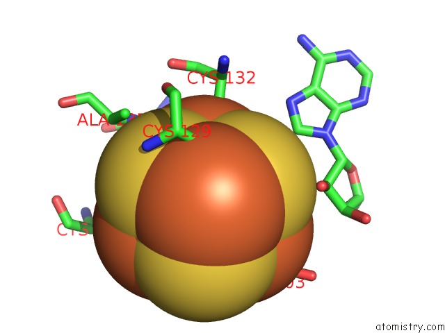
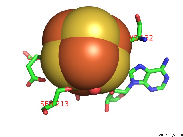
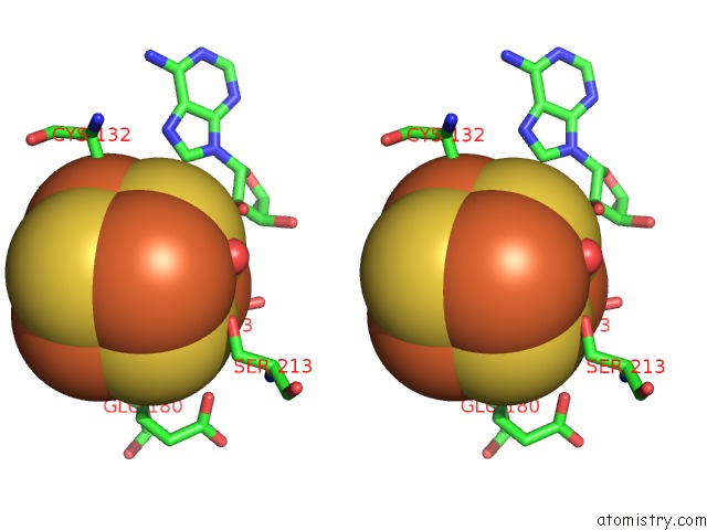
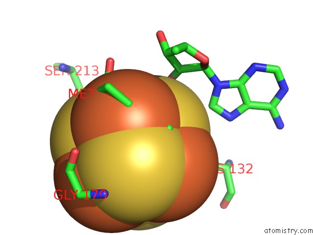
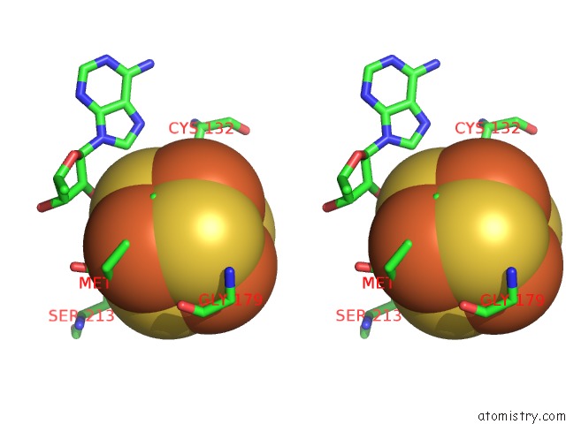
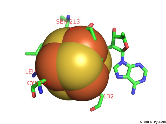
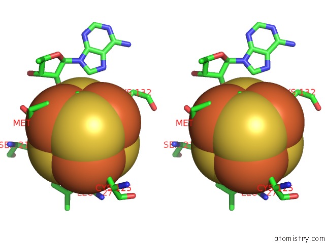
Iron binding site 1 out of 8 in 5hr7
Go back to
Iron binding site 1 out
of 8 in the X-Ray Crystal Structure of C118A Rlmn From Escherichia Coli with Cross-Linked in Vitro Transcribed Trna
Mono view
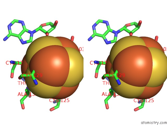
Stereo pair view
Mono view
Stereo pair view
A full contact list of Iron with other atoms in the Fe binding
site number 1 of X-Ray Crystal Structure of C118A Rlmn From Escherichia Coli with Cross-Linked in Vitro Transcribed Trna within 5.0Å range:
|
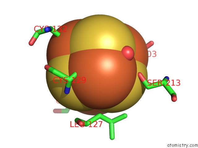
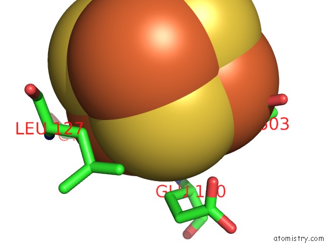
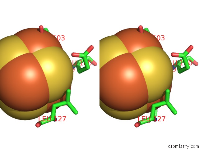
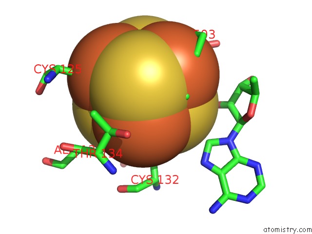
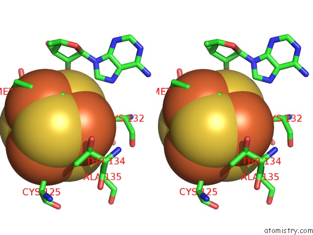
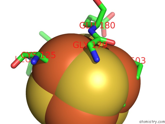
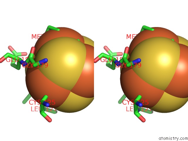
Iron binding site 2 out of 8 in 5hr7
Go back to
Iron binding site 2 out
of 8 in the X-Ray Crystal Structure of C118A Rlmn From Escherichia Coli with Cross-Linked in Vitro Transcribed Trna
Mono view
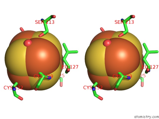
Stereo pair view
Mono view
Stereo pair view
A full contact list of Iron with other atoms in the Fe binding
site number 2 of X-Ray Crystal Structure of C118A Rlmn From Escherichia Coli with Cross-Linked in Vitro Transcribed Trna within 5.0Å range:
|
Iron binding site 3 out of 8 in 5hr7
Go back to
Iron binding site 3 out
of 8 in the X-Ray Crystal Structure of C118A Rlmn From Escherichia Coli with Cross-Linked in Vitro Transcribed Trna
Mono view
Stereo pair view
Mono view
Stereo pair view
A full contact list of Iron with other atoms in the Fe binding
site number 3 of X-Ray Crystal Structure of C118A Rlmn From Escherichia Coli with Cross-Linked in Vitro Transcribed Trna within 5.0Å range:
|
Iron binding site 4 out of 8 in 5hr7
Go back to
Iron binding site 4 out
of 8 in the X-Ray Crystal Structure of C118A Rlmn From Escherichia Coli with Cross-Linked in Vitro Transcribed Trna
Mono view
Stereo pair view
Mono view
Stereo pair view
A full contact list of Iron with other atoms in the Fe binding
site number 4 of X-Ray Crystal Structure of C118A Rlmn From Escherichia Coli with Cross-Linked in Vitro Transcribed Trna within 5.0Å range:
|
Iron binding site 5 out of 8 in 5hr7
Go back to
Iron binding site 5 out
of 8 in the X-Ray Crystal Structure of C118A Rlmn From Escherichia Coli with Cross-Linked in Vitro Transcribed Trna
Mono view
Stereo pair view
Mono view
Stereo pair view
A full contact list of Iron with other atoms in the Fe binding
site number 5 of X-Ray Crystal Structure of C118A Rlmn From Escherichia Coli with Cross-Linked in Vitro Transcribed Trna within 5.0Å range:
|
Iron binding site 6 out of 8 in 5hr7
Go back to
Iron binding site 6 out
of 8 in the X-Ray Crystal Structure of C118A Rlmn From Escherichia Coli with Cross-Linked in Vitro Transcribed Trna
Mono view
Stereo pair view
Mono view
Stereo pair view
A full contact list of Iron with other atoms in the Fe binding
site number 6 of X-Ray Crystal Structure of C118A Rlmn From Escherichia Coli with Cross-Linked in Vitro Transcribed Trna within 5.0Å range:
|
Iron binding site 7 out of 8 in 5hr7
Go back to
Iron binding site 7 out
of 8 in the X-Ray Crystal Structure of C118A Rlmn From Escherichia Coli with Cross-Linked in Vitro Transcribed Trna
Mono view
Stereo pair view
Mono view
Stereo pair view
A full contact list of Iron with other atoms in the Fe binding
site number 7 of X-Ray Crystal Structure of C118A Rlmn From Escherichia Coli with Cross-Linked in Vitro Transcribed Trna within 5.0Å range:
|
Iron binding site 8 out of 8 in 5hr7
Go back to
Iron binding site 8 out
of 8 in the X-Ray Crystal Structure of C118A Rlmn From Escherichia Coli with Cross-Linked in Vitro Transcribed Trna
Mono view
Stereo pair view
Mono view
Stereo pair view
A full contact list of Iron with other atoms in the Fe binding
site number 8 of X-Ray Crystal Structure of C118A Rlmn From Escherichia Coli with Cross-Linked in Vitro Transcribed Trna within 5.0Å range:
|
Reference:
E.L.Schwalm,
T.L.Grove,
S.J.Booker,
A.K.Boal.
Crystallographic Capture of A Radical S-Adenosylmethionine Enzyme in the Act of Modifying Trna. Science V. 352 309 2016.
ISSN: ESSN 1095-9203
PubMed: 27081063
DOI: 10.1126/SCIENCE.AAD5367
Page generated: Tue Aug 5 21:56:22 2025
ISSN: ESSN 1095-9203
PubMed: 27081063
DOI: 10.1126/SCIENCE.AAD5367
Last articles
I in 5W1HI in 5W1I
I in 5W0M
I in 5W0N
I in 5W0B
I in 5W0J
I in 5V65
I in 5VTE
I in 5VF1
I in 5VQ5