Iron »
PDB 5ibf-5j84 »
5ir6 »
Iron in PDB 5ir6: The Structure of Bd Oxidase From Geobacillus Thermodenitrificans
Protein crystallography data
The structure of The Structure of Bd Oxidase From Geobacillus Thermodenitrificans, PDB code: 5ir6
was solved by
S.Safarian,
H.Mueller,
C.Rajendran,
J.Preu,
S.Ovchinnikov,
T.Kusumoto,
T.Hirose,
J.Langer,
J.Sakamoto,
H.Michel,
with X-Ray Crystallography technique. A brief refinement statistics is given in the table below:
| Resolution Low / High (Å) | 20.00 / 3.80 |
| Space group | P 21 21 21 |
| Cell size a, b, c (Å), α, β, γ (°) | 110.060, 120.860, 122.720, 90.00, 90.00, 90.00 |
| R / Rfree (%) | 30.3 / 32.5 |
Iron Binding Sites:
The binding sites of Iron atom in the The Structure of Bd Oxidase From Geobacillus Thermodenitrificans
(pdb code 5ir6). This binding sites where shown within
5.0 Angstroms radius around Iron atom.
In total 3 binding sites of Iron where determined in the The Structure of Bd Oxidase From Geobacillus Thermodenitrificans, PDB code: 5ir6:
Jump to Iron binding site number: 1; 2; 3;
In total 3 binding sites of Iron where determined in the The Structure of Bd Oxidase From Geobacillus Thermodenitrificans, PDB code: 5ir6:
Jump to Iron binding site number: 1; 2; 3;
Iron binding site 1 out of 3 in 5ir6
Go back to
Iron binding site 1 out
of 3 in the The Structure of Bd Oxidase From Geobacillus Thermodenitrificans

Mono view

Stereo pair view
Mono view
Stereo pair view
A full contact list of Iron with other atoms in the Fe binding
site number 1 of The Structure of Bd Oxidase From Geobacillus Thermodenitrificans within 5.0Å range:
|
Iron binding site 2 out of 3 in 5ir6
Go back to
Iron binding site 2 out
of 3 in the The Structure of Bd Oxidase From Geobacillus Thermodenitrificans

Mono view
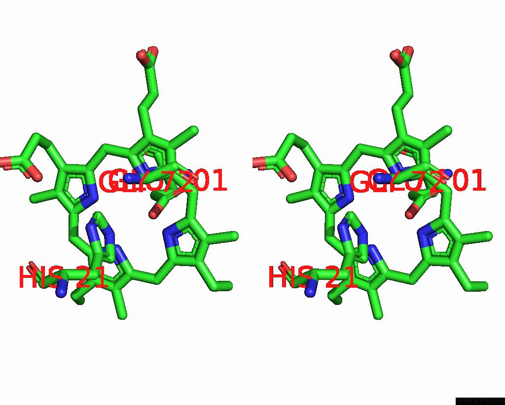
Stereo pair view
Mono view
Stereo pair view
A full contact list of Iron with other atoms in the Fe binding
site number 2 of The Structure of Bd Oxidase From Geobacillus Thermodenitrificans within 5.0Å range:
|
Iron binding site 3 out of 3 in 5ir6
Go back to
Iron binding site 3 out
of 3 in the The Structure of Bd Oxidase From Geobacillus Thermodenitrificans

Mono view

Stereo pair view
Mono view
Stereo pair view
A full contact list of Iron with other atoms in the Fe binding
site number 3 of The Structure of Bd Oxidase From Geobacillus Thermodenitrificans within 5.0Å range:
|
Reference:
S.Safarian,
C.Rajendran,
H.Muller,
J.Preu,
J.D.Langer,
S.Ovchinnikov,
T.Hirose,
T.Kusumoto,
J.Sakamoto,
H.Michel.
Structure of A Bd Oxidase Indicates Similar Mechanisms For Membrane-Integrated Oxygen Reductases. Science V. 352 583 2016.
ISSN: ESSN 1095-9203
PubMed: 27126043
DOI: 10.1126/SCIENCE.AAF2477
Page generated: Tue Aug 5 22:02:41 2025
ISSN: ESSN 1095-9203
PubMed: 27126043
DOI: 10.1126/SCIENCE.AAF2477
Last articles
Na in 4PZ2Na in 4Q1J
Na in 4Q1H
Na in 4PYQ
Na in 4Q04
Na in 4PY9
Na in 4PYK
Na in 4PYJ
Na in 4PYI
Na in 4PXL