Iron »
PDB 5ibf-5j84 »
5ixz »
Iron in PDB 5ixz: Crystal Structure of the Signaling Protein Complex 5
Protein crystallography data
The structure of Crystal Structure of the Signaling Protein Complex 5, PDB code: 5ixz
was solved by
V.Kumar,
F.Van Den Akker,
with X-Ray Crystallography technique. A brief refinement statistics is given in the table below:
| Resolution Low / High (Å) | 38.98 / 2.40 |
| Space group | P 21 3 |
| Cell size a, b, c (Å), α, β, γ (°) | 123.267, 123.267, 123.267, 90.00, 90.00, 90.00 |
| R / Rfree (%) | 14.9 / 17.7 |
Iron Binding Sites:
The binding sites of Iron atom in the Crystal Structure of the Signaling Protein Complex 5
(pdb code 5ixz). This binding sites where shown within
5.0 Angstroms radius around Iron atom.
In total 2 binding sites of Iron where determined in the Crystal Structure of the Signaling Protein Complex 5, PDB code: 5ixz:
Jump to Iron binding site number: 1; 2;
In total 2 binding sites of Iron where determined in the Crystal Structure of the Signaling Protein Complex 5, PDB code: 5ixz:
Jump to Iron binding site number: 1; 2;
Iron binding site 1 out of 2 in 5ixz
Go back to
Iron binding site 1 out
of 2 in the Crystal Structure of the Signaling Protein Complex 5
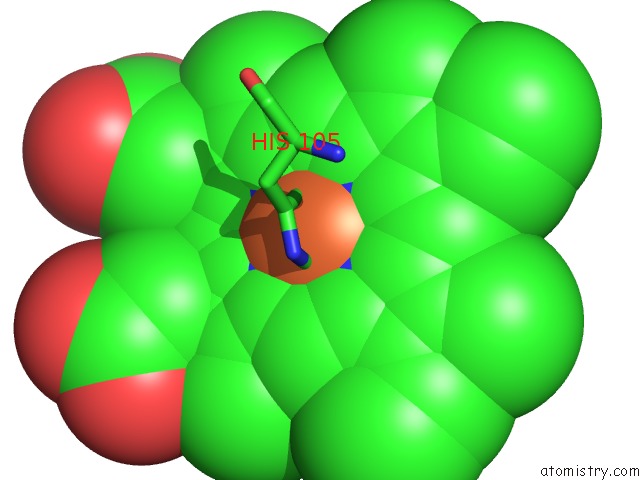
Mono view
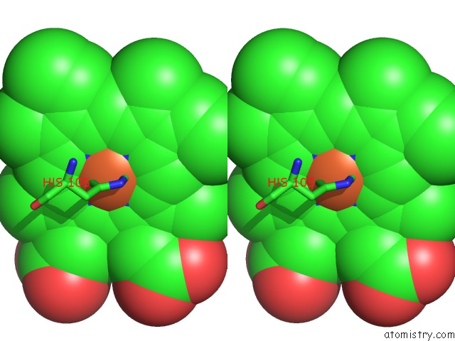
Stereo pair view
Mono view
Stereo pair view
A full contact list of Iron with other atoms in the Fe binding
site number 1 of Crystal Structure of the Signaling Protein Complex 5 within 5.0Å range:
|
Iron binding site 2 out of 2 in 5ixz
Go back to
Iron binding site 2 out
of 2 in the Crystal Structure of the Signaling Protein Complex 5
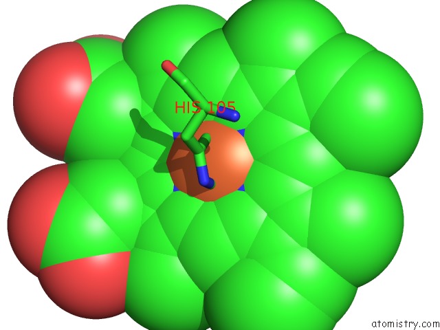
Mono view
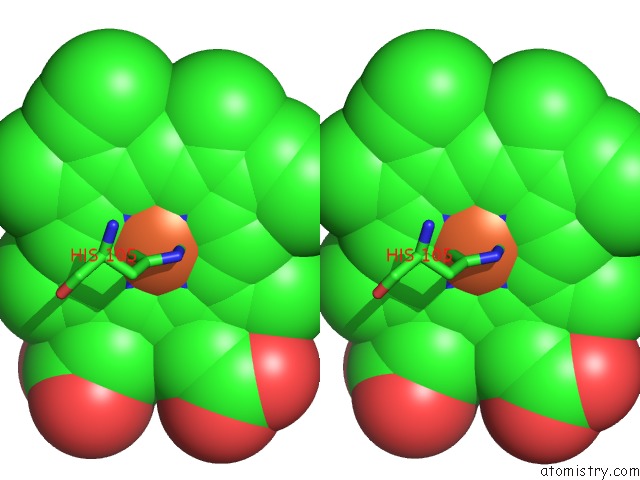
Stereo pair view
Mono view
Stereo pair view
A full contact list of Iron with other atoms in the Fe binding
site number 2 of Crystal Structure of the Signaling Protein Complex 5 within 5.0Å range:
|
Reference:
V.Kumar,
F.Van Den Akker.
Crystal Structure of the Signaling Protein Complex 5 To Be Published.
Page generated: Tue Aug 5 22:04:35 2025
Last articles
Na in 4PO9Na in 4PNZ
Na in 4PNC
Na in 4PMR
Na in 4PMF
Na in 4PLH
Na in 4PJH
Na in 4PME
Na in 4PHI
Na in 4PK0