Iron »
PDB 5kts-5lg8 »
5l1o »
Iron in PDB 5l1o: X-Ray Structure of Cytochrome P450 Pntm with Pentalenolactone F
Enzymatic activity of X-Ray Structure of Cytochrome P450 Pntm with Pentalenolactone F
All present enzymatic activity of X-Ray Structure of Cytochrome P450 Pntm with Pentalenolactone F:
1.14.19.8;
1.14.19.8;
Protein crystallography data
The structure of X-Ray Structure of Cytochrome P450 Pntm with Pentalenolactone F, PDB code: 5l1o
was solved by
L.Duan,
G.Jogl,
D.E.Cane,
with X-Ray Crystallography technique. A brief refinement statistics is given in the table below:
| Resolution Low / High (Å) | 45.79 / 2.03 |
| Space group | P 21 21 2 |
| Cell size a, b, c (Å), α, β, γ (°) | 44.746, 164.475, 83.289, 90.00, 90.00, 90.00 |
| R / Rfree (%) | 14.3 / 17.8 |
Iron Binding Sites:
The binding sites of Iron atom in the X-Ray Structure of Cytochrome P450 Pntm with Pentalenolactone F
(pdb code 5l1o). This binding sites where shown within
5.0 Angstroms radius around Iron atom.
In total only one binding site of Iron was determined in the X-Ray Structure of Cytochrome P450 Pntm with Pentalenolactone F, PDB code: 5l1o:
In total only one binding site of Iron was determined in the X-Ray Structure of Cytochrome P450 Pntm with Pentalenolactone F, PDB code: 5l1o:
Iron binding site 1 out of 1 in 5l1o
Go back to
Iron binding site 1 out
of 1 in the X-Ray Structure of Cytochrome P450 Pntm with Pentalenolactone F
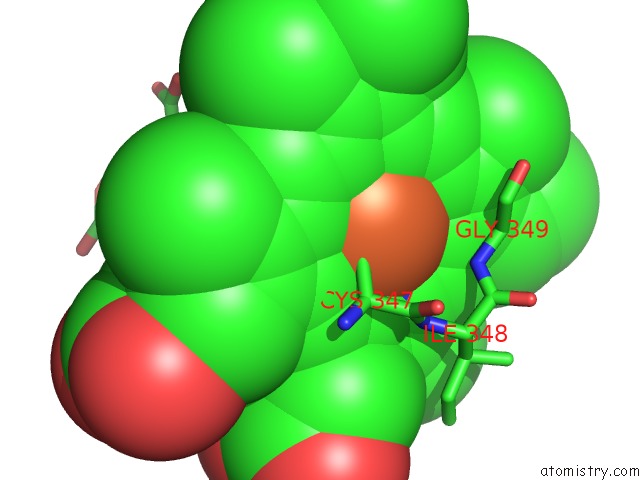
Mono view
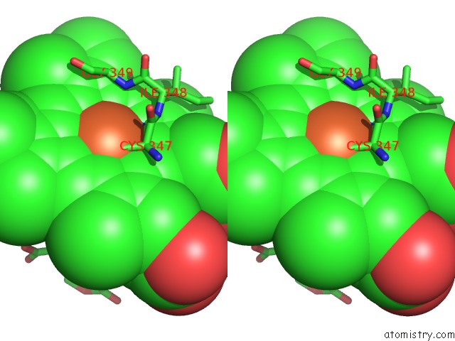
Stereo pair view
Mono view
Stereo pair view
A full contact list of Iron with other atoms in the Fe binding
site number 1 of X-Ray Structure of Cytochrome P450 Pntm with Pentalenolactone F within 5.0Å range:
|
Reference:
L.Duan,
G.Jogl,
D.E.Cane.
The Cytochrome P450-Catalyzed Oxidative Rearrangement in the Final Step of Pentalenolactone Biosynthesis: Substrate Structure Determines Mechanism. J.Am.Chem.Soc. V. 138 12678 2016.
ISSN: ESSN 1520-5126
PubMed: 27588339
DOI: 10.1021/JACS.6B08610
Page generated: Tue Aug 5 22:56:10 2025
ISSN: ESSN 1520-5126
PubMed: 27588339
DOI: 10.1021/JACS.6B08610
Last articles
Mn in 9LJUMn in 9LJW
Mn in 9LJS
Mn in 9LJR
Mn in 9LJT
Mn in 9LJV
Mg in 9UA2
Mg in 9R96
Mg in 9VM1
Mg in 9P01