Iron »
PDB 5li6-5m2g »
5m21 »
Iron in PDB 5m21: Crystal Structure of Hydroquinone 1,2-Dioxygenase From Sphingomonas Sp. TTNP3 with 4-Hydroxybenzoate Bound
Protein crystallography data
The structure of Crystal Structure of Hydroquinone 1,2-Dioxygenase From Sphingomonas Sp. TTNP3 with 4-Hydroxybenzoate Bound, PDB code: 5m21
was solved by
M.Ferraroni,
S.Da Vela,
A.Scozzafava,
B.Kolvenbach,
P.F.X.Corvini,
with X-Ray Crystallography technique. A brief refinement statistics is given in the table below:
| Resolution Low / High (Å) | 29.80 / 1.99 |
| Space group | P 1 21 1 |
| Cell size a, b, c (Å), α, β, γ (°) | 88.782, 124.869, 92.371, 90.00, 105.15, 90.00 |
| R / Rfree (%) | 17.9 / 24.2 |
Iron Binding Sites:
The binding sites of Iron atom in the Crystal Structure of Hydroquinone 1,2-Dioxygenase From Sphingomonas Sp. TTNP3 with 4-Hydroxybenzoate Bound
(pdb code 5m21). This binding sites where shown within
5.0 Angstroms radius around Iron atom.
In total 4 binding sites of Iron where determined in the Crystal Structure of Hydroquinone 1,2-Dioxygenase From Sphingomonas Sp. TTNP3 with 4-Hydroxybenzoate Bound, PDB code: 5m21:
Jump to Iron binding site number: 1; 2; 3; 4;
In total 4 binding sites of Iron where determined in the Crystal Structure of Hydroquinone 1,2-Dioxygenase From Sphingomonas Sp. TTNP3 with 4-Hydroxybenzoate Bound, PDB code: 5m21:
Jump to Iron binding site number: 1; 2; 3; 4;
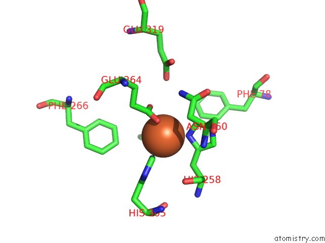
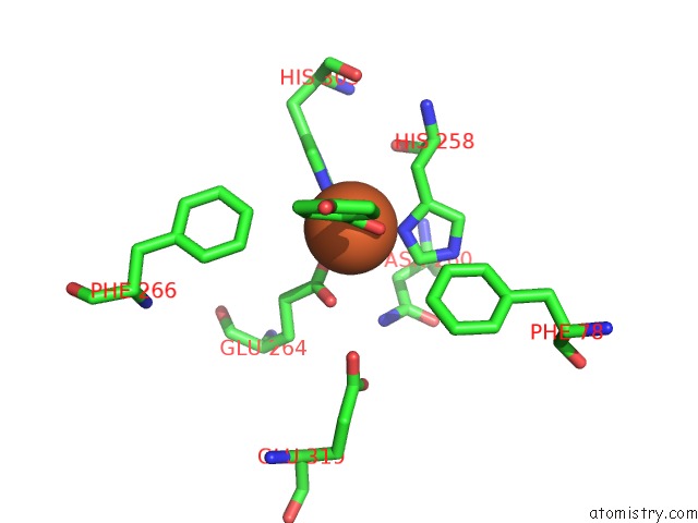
Iron binding site 1 out of 4 in 5m21
Go back to
Iron binding site 1 out
of 4 in the Crystal Structure of Hydroquinone 1,2-Dioxygenase From Sphingomonas Sp. TTNP3 with 4-Hydroxybenzoate Bound
Mono view
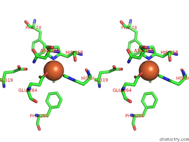
Stereo pair view
Mono view
Stereo pair view
A full contact list of Iron with other atoms in the Fe binding
site number 1 of Crystal Structure of Hydroquinone 1,2-Dioxygenase From Sphingomonas Sp. TTNP3 with 4-Hydroxybenzoate Bound within 5.0Å range:
|
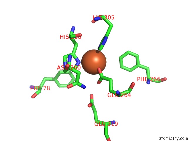
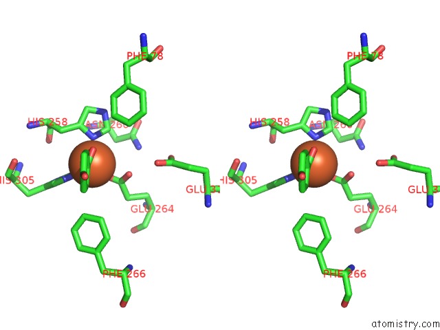
Iron binding site 2 out of 4 in 5m21
Go back to
Iron binding site 2 out
of 4 in the Crystal Structure of Hydroquinone 1,2-Dioxygenase From Sphingomonas Sp. TTNP3 with 4-Hydroxybenzoate Bound
Mono view
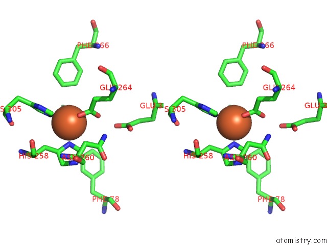
Stereo pair view
Mono view
Stereo pair view
A full contact list of Iron with other atoms in the Fe binding
site number 2 of Crystal Structure of Hydroquinone 1,2-Dioxygenase From Sphingomonas Sp. TTNP3 with 4-Hydroxybenzoate Bound within 5.0Å range:
|
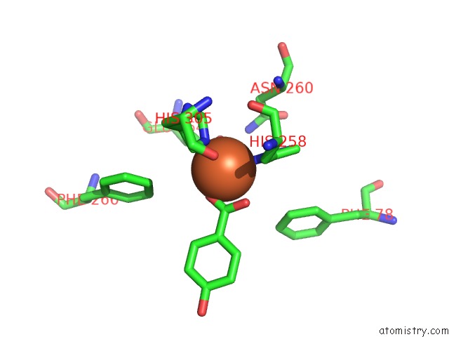
Iron binding site 3 out of 4 in 5m21
Go back to
Iron binding site 3 out
of 4 in the Crystal Structure of Hydroquinone 1,2-Dioxygenase From Sphingomonas Sp. TTNP3 with 4-Hydroxybenzoate Bound
Mono view
Stereo pair view
Mono view
Stereo pair view
A full contact list of Iron with other atoms in the Fe binding
site number 3 of Crystal Structure of Hydroquinone 1,2-Dioxygenase From Sphingomonas Sp. TTNP3 with 4-Hydroxybenzoate Bound within 5.0Å range:
|
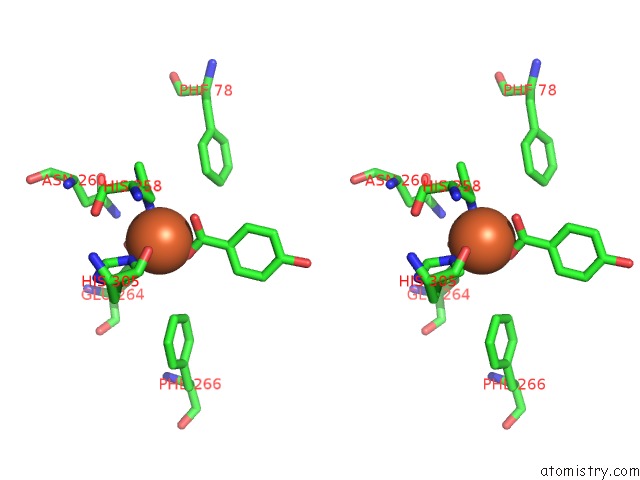
Iron binding site 4 out of 4 in 5m21
Go back to
Iron binding site 4 out
of 4 in the Crystal Structure of Hydroquinone 1,2-Dioxygenase From Sphingomonas Sp. TTNP3 with 4-Hydroxybenzoate Bound
Mono view
Stereo pair view
Mono view
Stereo pair view
A full contact list of Iron with other atoms in the Fe binding
site number 4 of Crystal Structure of Hydroquinone 1,2-Dioxygenase From Sphingomonas Sp. TTNP3 with 4-Hydroxybenzoate Bound within 5.0Å range:
|
Reference:
M.Ferraroni,
S.Da Vela,
B.A.Kolvenbach,
P.F.Corvini,
A.Scozzafava.
The Crystal Structures of Native Hydroquinone 1,2-Dioxygenase From Sphingomonas Sp. TTNP3 and of Substrate and Inhibitor Complexes. Biochim. Biophys. Acta V.1865 520 2017.
ISSN: ISSN 0006-3002
PubMed: 28232026
DOI: 10.1016/J.BBAPAP.2017.02.013
Page generated: Tue Aug 6 04:56:12 2024
ISSN: ISSN 0006-3002
PubMed: 28232026
DOI: 10.1016/J.BBAPAP.2017.02.013
Last articles
Fe in 2YXOFe in 2YRS
Fe in 2YXC
Fe in 2YNM
Fe in 2YVJ
Fe in 2YP1
Fe in 2YU2
Fe in 2YU1
Fe in 2YQB
Fe in 2YOO