Iron »
PDB 5m3l-5mkq »
5m6j »
Iron in PDB 5m6j: Crystal Structure of Nitrophorin 7 E27V Mutant From Rhodnius Prolixus
Enzymatic activity of Crystal Structure of Nitrophorin 7 E27V Mutant From Rhodnius Prolixus
All present enzymatic activity of Crystal Structure of Nitrophorin 7 E27V Mutant From Rhodnius Prolixus:
1.7.6.1;
1.7.6.1;
Protein crystallography data
The structure of Crystal Structure of Nitrophorin 7 E27V Mutant From Rhodnius Prolixus, PDB code: 5m6j
was solved by
H.Ogata,
with X-Ray Crystallography technique. A brief refinement statistics is given in the table below:
| Resolution Low / High (Å) | 34.63 / 1.70 |
| Space group | P 1 21 1 |
| Cell size a, b, c (Å), α, β, γ (°) | 38.228, 66.885, 38.714, 90.00, 116.57, 90.00 |
| R / Rfree (%) | 16.1 / 21.3 |
Iron Binding Sites:
The binding sites of Iron atom in the Crystal Structure of Nitrophorin 7 E27V Mutant From Rhodnius Prolixus
(pdb code 5m6j). This binding sites where shown within
5.0 Angstroms radius around Iron atom.
In total only one binding site of Iron was determined in the Crystal Structure of Nitrophorin 7 E27V Mutant From Rhodnius Prolixus, PDB code: 5m6j:
In total only one binding site of Iron was determined in the Crystal Structure of Nitrophorin 7 E27V Mutant From Rhodnius Prolixus, PDB code: 5m6j:
Iron binding site 1 out of 1 in 5m6j
Go back to
Iron binding site 1 out
of 1 in the Crystal Structure of Nitrophorin 7 E27V Mutant From Rhodnius Prolixus
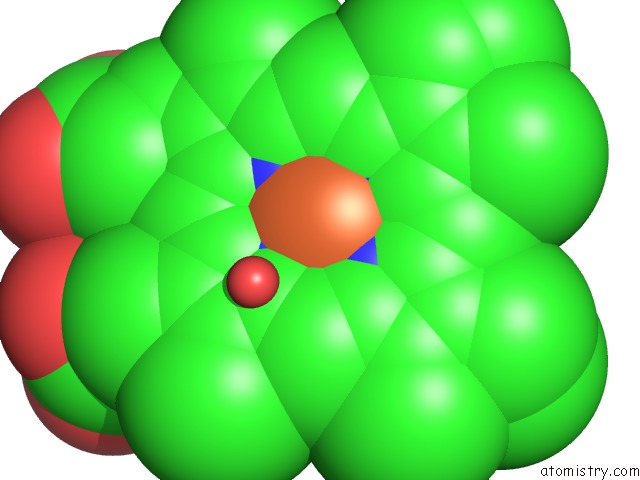
Mono view
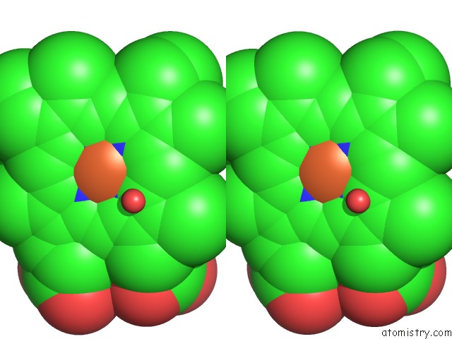
Stereo pair view
Mono view
Stereo pair view
A full contact list of Iron with other atoms in the Fe binding
site number 1 of Crystal Structure of Nitrophorin 7 E27V Mutant From Rhodnius Prolixus within 5.0Å range:
|
Reference:
S.Abbruzzetti,
A.Allegri,
A.Bidon-Chanal,
H.Ogata,
G.Soavi,
G.Cerullo,
S.Bruno,
C.Montali,
F.J.Luque,
C.Viappiani.
Electrostatic Tuning of the Ligand Binding Mechanism By GLU27 in Nitrophorin 7. Sci Rep V. 8 10855 2018.
ISSN: ESSN 2045-2322
PubMed: 30022039
DOI: 10.1038/S41598-018-29182-3
Page generated: Tue Aug 5 23:32:17 2025
ISSN: ESSN 2045-2322
PubMed: 30022039
DOI: 10.1038/S41598-018-29182-3
Last articles
Mg in 4E7ZMg in 4E6M
Mg in 4E7S
Mg in 4E7P
Mg in 4E7O
Mg in 4E5T
Mg in 4E6N
Mg in 4E6E
Mg in 4E4P
Mg in 4E4F