Iron »
PDB 5mms-5o10 »
5nc0 »
Iron in PDB 5nc0: The 0.91 A Resolution Structure of the L16G Mutant of Cytochrome C Prime From Alcaligenes Xylosoxidans, Complexed with Nitric Oxide
Protein crystallography data
The structure of The 0.91 A Resolution Structure of the L16G Mutant of Cytochrome C Prime From Alcaligenes Xylosoxidans, Complexed with Nitric Oxide, PDB code: 5nc0
was solved by
R.Strange,
M.Hough,
S.Antonyuk,
N.Rustage,
with X-Ray Crystallography technique. A brief refinement statistics is given in the table below:
| Resolution Low / High (Å) | 44.07 / 0.91 |
| Space group | P 65 2 2 |
| Cell size a, b, c (Å), α, β, γ (°) | 52.430, 52.430, 183.250, 90.00, 90.00, 120.00 |
| R / Rfree (%) | 12.5 / 13.2 |
Iron Binding Sites:
The binding sites of Iron atom in the The 0.91 A Resolution Structure of the L16G Mutant of Cytochrome C Prime From Alcaligenes Xylosoxidans, Complexed with Nitric Oxide
(pdb code 5nc0). This binding sites where shown within
5.0 Angstroms radius around Iron atom.
In total only one binding site of Iron was determined in the The 0.91 A Resolution Structure of the L16G Mutant of Cytochrome C Prime From Alcaligenes Xylosoxidans, Complexed with Nitric Oxide, PDB code: 5nc0:
In total only one binding site of Iron was determined in the The 0.91 A Resolution Structure of the L16G Mutant of Cytochrome C Prime From Alcaligenes Xylosoxidans, Complexed with Nitric Oxide, PDB code: 5nc0:
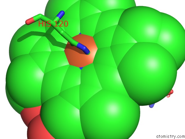
Iron binding site 1 out of 1 in 5nc0
Go back to
Iron binding site 1 out
of 1 in the The 0.91 A Resolution Structure of the L16G Mutant of Cytochrome C Prime From Alcaligenes Xylosoxidans, Complexed with Nitric Oxide
Mono view

Stereo pair view
Mono view
Stereo pair view
A full contact list of Iron with other atoms in the Fe binding
site number 1 of The 0.91 A Resolution Structure of the L16G Mutant of Cytochrome C Prime From Alcaligenes Xylosoxidans, Complexed with Nitric Oxide within 5.0Å range:
|
Reference:
Z.N.Nilsson,
B.L.Mandella,
K.Sen,
D.Kekilli,
M.A.Hough,
P.Moenne-Loccoz,
R.W.Strange,
C.R.Andrew.
Distinguishing Nitro Vs Nitrito Coordination in Cytochrome C' Using Vibrational Spectroscopy and Density Functional Theory. Inorg Chem V. 56 13205 2017.
ISSN: ISSN 1520-510X
PubMed: 29053273
DOI: 10.1021/ACS.INORGCHEM.7B01945
Page generated: Tue Aug 6 06:07:57 2024
ISSN: ISSN 1520-510X
PubMed: 29053273
DOI: 10.1021/ACS.INORGCHEM.7B01945
Last articles
Zn in 9MJ5Zn in 9HNW
Zn in 9G0L
Zn in 9FNE
Zn in 9DZN
Zn in 9E0I
Zn in 9D32
Zn in 9DAK
Zn in 8ZXC
Zn in 8ZUF