Iron »
PDB 5sx3-5tia »
5t8z »
Iron in PDB 5t8z: Crystal Structure of A Peptide Deformylase From Burkholderia Multivorans in Complex with Actinonin
Enzymatic activity of Crystal Structure of A Peptide Deformylase From Burkholderia Multivorans in Complex with Actinonin
All present enzymatic activity of Crystal Structure of A Peptide Deformylase From Burkholderia Multivorans in Complex with Actinonin:
3.5.1.88;
3.5.1.88;
Protein crystallography data
The structure of Crystal Structure of A Peptide Deformylase From Burkholderia Multivorans in Complex with Actinonin, PDB code: 5t8z
was solved by
Seattle Structural Genomics Center For Infectious Disease (Ssgcid),
with X-Ray Crystallography technique. A brief refinement statistics is given in the table below:
| Resolution Low / High (Å) | 39.02 / 1.85 |
| Space group | I 2 2 2 |
| Cell size a, b, c (Å), α, β, γ (°) | 39.120, 70.540, 140.510, 90.00, 90.00, 90.00 |
| R / Rfree (%) | 16.5 / 20.3 |
Iron Binding Sites:
The binding sites of Iron atom in the Crystal Structure of A Peptide Deformylase From Burkholderia Multivorans in Complex with Actinonin
(pdb code 5t8z). This binding sites where shown within
5.0 Angstroms radius around Iron atom.
In total only one binding site of Iron was determined in the Crystal Structure of A Peptide Deformylase From Burkholderia Multivorans in Complex with Actinonin, PDB code: 5t8z:
In total only one binding site of Iron was determined in the Crystal Structure of A Peptide Deformylase From Burkholderia Multivorans in Complex with Actinonin, PDB code: 5t8z:
Iron binding site 1 out of 1 in 5t8z
Go back to
Iron binding site 1 out
of 1 in the Crystal Structure of A Peptide Deformylase From Burkholderia Multivorans in Complex with Actinonin
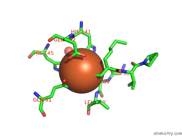
Mono view
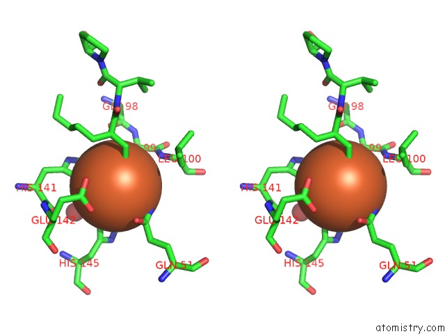
Stereo pair view
Mono view
Stereo pair view
A full contact list of Iron with other atoms in the Fe binding
site number 1 of Crystal Structure of A Peptide Deformylase From Burkholderia Multivorans in Complex with Actinonin within 5.0Å range:
|
Reference:
R.M.Irwin,
J.Abendroth,
S.J.Mayclin,
D.D.Lorimer,
T.E.Edwards.
Crystal Structure of A Peptide Deformylase From Burkholderia Multivorans in Complex with Actinonin To Be Published.
Page generated: Wed Aug 6 01:29:47 2025
Last articles
Xe in 7B2CXe in 7SZG
Xe in 7B2H
Xe in 7SHW
Xe in 6AYK
Xe in 6QII
Xe in 6L9X
Xe in 5NSW
Xe in 4ZZC
Xe in 6FY9