Iron »
PDB 5vux-5whs »
5vxt »
Iron in PDB 5vxt: Crystal Structure of Catechol 1,2-Dioxygenase From Burkholderia Ambifaria
Protein crystallography data
The structure of Crystal Structure of Catechol 1,2-Dioxygenase From Burkholderia Ambifaria, PDB code: 5vxt
was solved by
Seattle Structural Genomics Center For Infectious Disease (Ssgcid),
with X-Ray Crystallography technique. A brief refinement statistics is given in the table below:
| Resolution Low / High (Å) | 47.81 / 1.75 |
| Space group | P 1 21 1 |
| Cell size a, b, c (Å), α, β, γ (°) | 66.080, 52.200, 105.190, 90.00, 106.62, 90.00 |
| R / Rfree (%) | 18.6 / 22.4 |
Other elements in 5vxt:
The structure of Crystal Structure of Catechol 1,2-Dioxygenase From Burkholderia Ambifaria also contains other interesting chemical elements:
| Chlorine | (Cl) | 1 atom |
Iron Binding Sites:
The binding sites of Iron atom in the Crystal Structure of Catechol 1,2-Dioxygenase From Burkholderia Ambifaria
(pdb code 5vxt). This binding sites where shown within
5.0 Angstroms radius around Iron atom.
In total 2 binding sites of Iron where determined in the Crystal Structure of Catechol 1,2-Dioxygenase From Burkholderia Ambifaria, PDB code: 5vxt:
Jump to Iron binding site number: 1; 2;
In total 2 binding sites of Iron where determined in the Crystal Structure of Catechol 1,2-Dioxygenase From Burkholderia Ambifaria, PDB code: 5vxt:
Jump to Iron binding site number: 1; 2;
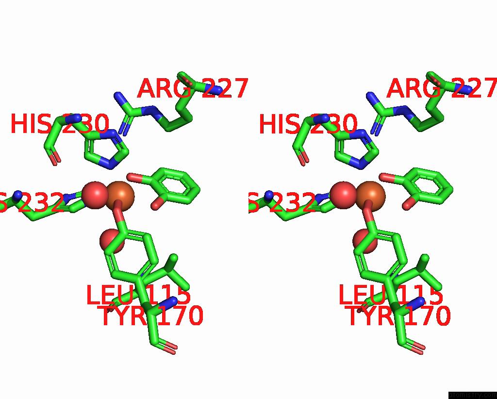
Iron binding site 1 out of 2 in 5vxt
Go back to
Iron binding site 1 out
of 2 in the Crystal Structure of Catechol 1,2-Dioxygenase From Burkholderia Ambifaria
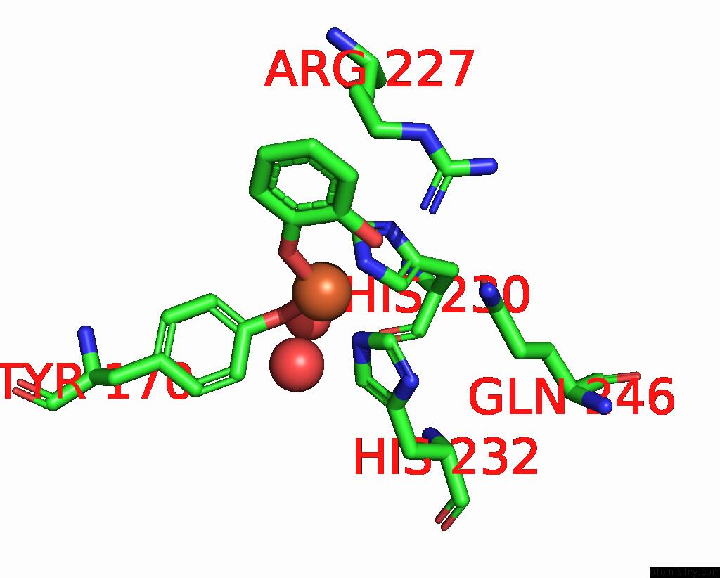
Mono view

Stereo pair view
Mono view
Stereo pair view
A full contact list of Iron with other atoms in the Fe binding
site number 1 of Crystal Structure of Catechol 1,2-Dioxygenase From Burkholderia Ambifaria within 5.0Å range:
|
Iron binding site 2 out of 2 in 5vxt
Go back to
Iron binding site 2 out
of 2 in the Crystal Structure of Catechol 1,2-Dioxygenase From Burkholderia Ambifaria

Mono view
Stereo pair view
Mono view
Stereo pair view
A full contact list of Iron with other atoms in the Fe binding
site number 2 of Crystal Structure of Catechol 1,2-Dioxygenase From Burkholderia Ambifaria within 5.0Å range:
|
Reference:
J.N.Phan,
D.M.Dranow,
D.Lorimer,
T.E.Edwards.
Crystal Structure of Catechol 1,2-Dioxygenase From Burkholderia Ambifaria To Be Published.
Page generated: Wed Aug 6 02:27:21 2025
Last articles
K in 6P9VK in 6P0Y
K in 6OZI
K in 6P1M
K in 6OZJ
K in 6OZR
K in 6OXD
K in 6OF8
K in 6OSR
K in 6OVL