Iron »
PDB 5xdh-5xy4 »
5xe1 »
Iron in PDB 5xe1: Crystal Structure of the Indoleamine 2,3-Dioxygenagse 1 (IDO1) Complexed with INCB14943
Enzymatic activity of Crystal Structure of the Indoleamine 2,3-Dioxygenagse 1 (IDO1) Complexed with INCB14943
All present enzymatic activity of Crystal Structure of the Indoleamine 2,3-Dioxygenagse 1 (IDO1) Complexed with INCB14943:
1.13.11.52;
1.13.11.52;
Protein crystallography data
The structure of Crystal Structure of the Indoleamine 2,3-Dioxygenagse 1 (IDO1) Complexed with INCB14943, PDB code: 5xe1
was solved by
J.Xu,
U.Wu,
J.Liu,
with X-Ray Crystallography technique. A brief refinement statistics is given in the table below:
| Resolution Low / High (Å) | 25.00 / 3.20 |
| Space group | P 21 21 21 |
| Cell size a, b, c (Å), α, β, γ (°) | 87.050, 97.380, 128.470, 90.00, 90.00, 90.00 |
| R / Rfree (%) | 22.8 / 27.4 |
Other elements in 5xe1:
The structure of Crystal Structure of the Indoleamine 2,3-Dioxygenagse 1 (IDO1) Complexed with INCB14943 also contains other interesting chemical elements:
| Fluorine | (F) | 2 atoms |
| Chlorine | (Cl) | 2 atoms |
Iron Binding Sites:
The binding sites of Iron atom in the Crystal Structure of the Indoleamine 2,3-Dioxygenagse 1 (IDO1) Complexed with INCB14943
(pdb code 5xe1). This binding sites where shown within
5.0 Angstroms radius around Iron atom.
In total 2 binding sites of Iron where determined in the Crystal Structure of the Indoleamine 2,3-Dioxygenagse 1 (IDO1) Complexed with INCB14943, PDB code: 5xe1:
Jump to Iron binding site number: 1; 2;
In total 2 binding sites of Iron where determined in the Crystal Structure of the Indoleamine 2,3-Dioxygenagse 1 (IDO1) Complexed with INCB14943, PDB code: 5xe1:
Jump to Iron binding site number: 1; 2;
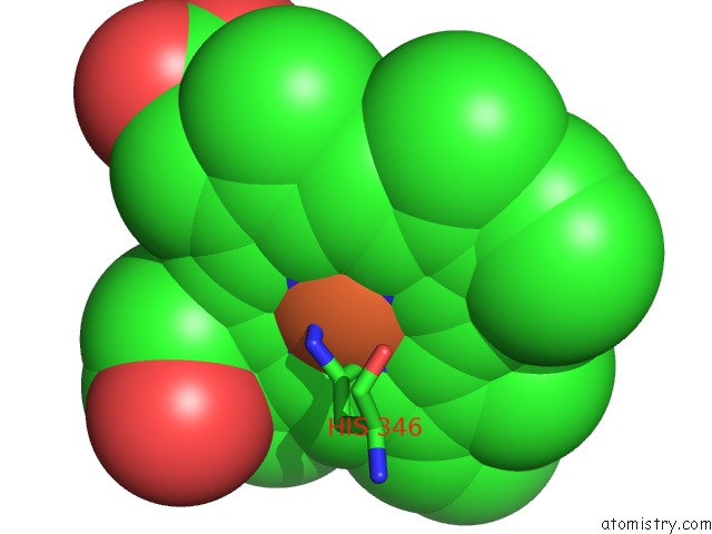
Iron binding site 1 out of 2 in 5xe1
Go back to
Iron binding site 1 out
of 2 in the Crystal Structure of the Indoleamine 2,3-Dioxygenagse 1 (IDO1) Complexed with INCB14943

Mono view

Stereo pair view
Mono view
Stereo pair view
A full contact list of Iron with other atoms in the Fe binding
site number 1 of Crystal Structure of the Indoleamine 2,3-Dioxygenagse 1 (IDO1) Complexed with INCB14943 within 5.0Å range:
|
Iron binding site 2 out of 2 in 5xe1
Go back to
Iron binding site 2 out
of 2 in the Crystal Structure of the Indoleamine 2,3-Dioxygenagse 1 (IDO1) Complexed with INCB14943
Mono view

Stereo pair view
Mono view
Stereo pair view
A full contact list of Iron with other atoms in the Fe binding
site number 2 of Crystal Structure of the Indoleamine 2,3-Dioxygenagse 1 (IDO1) Complexed with INCB14943 within 5.0Å range:
|
Reference:
Y.Wu,
T.Xu,
J.Liu,
K.Ding,
J.Xu.
Structural Insights Into the Binding Mechanism of IDO1 with Hydroxylamidine Based Inhibitor INCB14943 Biochem. Biophys. Res. V. 487 339 2017COMMUN..
ISSN: ESSN 1090-2104
PubMed: 28412361
DOI: 10.1016/J.BBRC.2017.04.061
Page generated: Tue Aug 6 11:39:25 2024
ISSN: ESSN 1090-2104
PubMed: 28412361
DOI: 10.1016/J.BBRC.2017.04.061
Last articles
F in 7MGEF in 7MFD
F in 7MEW
F in 7ME8
F in 7MCF
F in 7MDP
F in 7M8V
F in 7MCK
F in 7MAZ
F in 7MBO