Iron »
PDB 5zm9-6air »
5zm9 »
Iron in PDB 5zm9: Crystal Structure of Hexacoordinated Heme Protein From Anhydrobiotic Tardigrade at pH 7
Protein crystallography data
The structure of Crystal Structure of Hexacoordinated Heme Protein From Anhydrobiotic Tardigrade at pH 7, PDB code: 5zm9
was solved by
J.Kim,
Y.Fukuda,
T.Inoue,
with X-Ray Crystallography technique. A brief refinement statistics is given in the table below:
| Resolution Low / High (Å) | 18.89 / 2.70 |
| Space group | P 21 21 21 |
| Cell size a, b, c (Å), α, β, γ (°) | 36.312, 56.627, 66.362, 90.00, 90.00, 90.00 |
| R / Rfree (%) | 21.7 / 26.7 |
Other elements in 5zm9:
The structure of Crystal Structure of Hexacoordinated Heme Protein From Anhydrobiotic Tardigrade at pH 7 also contains other interesting chemical elements:
| Chlorine | (Cl) | 1 atom |
Iron Binding Sites:
The binding sites of Iron atom in the Crystal Structure of Hexacoordinated Heme Protein From Anhydrobiotic Tardigrade at pH 7
(pdb code 5zm9). This binding sites where shown within
5.0 Angstroms radius around Iron atom.
In total only one binding site of Iron was determined in the Crystal Structure of Hexacoordinated Heme Protein From Anhydrobiotic Tardigrade at pH 7, PDB code: 5zm9:
In total only one binding site of Iron was determined in the Crystal Structure of Hexacoordinated Heme Protein From Anhydrobiotic Tardigrade at pH 7, PDB code: 5zm9:
Iron binding site 1 out of 1 in 5zm9
Go back to
Iron binding site 1 out
of 1 in the Crystal Structure of Hexacoordinated Heme Protein From Anhydrobiotic Tardigrade at pH 7
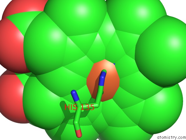
Mono view
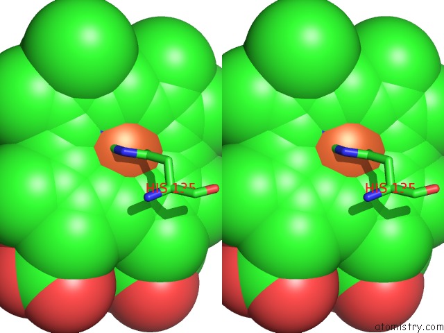
Stereo pair view
Mono view
Stereo pair view
A full contact list of Iron with other atoms in the Fe binding
site number 1 of Crystal Structure of Hexacoordinated Heme Protein From Anhydrobiotic Tardigrade at pH 7 within 5.0Å range:
|
Reference:
J.Kim,
Y.Fukuda,
T.Inoue.
Crystal Structure of Kumaglobin: A Hexacoordinated Heme Protein From An Anhydrobiotic Tardigrade, Ramazzottius Varieornatus. Febs J. V. 286 1287 2019.
ISSN: ISSN 1742-4658
PubMed: 30506636
DOI: 10.1111/FEBS.14713
Page generated: Wed Aug 6 03:37:47 2025
ISSN: ISSN 1742-4658
PubMed: 30506636
DOI: 10.1111/FEBS.14713
Last articles
I in 5JRFI in 5IQY
I in 5JTB
I in 5IJQ
I in 5JRV
I in 5JGP
I in 5IO8
I in 5IJS
I in 5IJW
I in 5GZH