Iron »
PDB 5zm9-6air »
6a2u »
Iron in PDB 6a2u: Crystal Structure of Gamma-Alpha Subunit Complex From Burkholderia Cepacia Fad Glucose Dehydrogenase
Enzymatic activity of Crystal Structure of Gamma-Alpha Subunit Complex From Burkholderia Cepacia Fad Glucose Dehydrogenase
All present enzymatic activity of Crystal Structure of Gamma-Alpha Subunit Complex From Burkholderia Cepacia Fad Glucose Dehydrogenase:
1.1.5.9;
1.1.5.9;
Protein crystallography data
The structure of Crystal Structure of Gamma-Alpha Subunit Complex From Burkholderia Cepacia Fad Glucose Dehydrogenase, PDB code: 6a2u
was solved by
H.Yoshida,
K.Kojima,
K.Yoshimatsu,
M.Shiota,
T.Yamazaki,
S.Ferri,
W.Tsugawa,
S.Kamitori,
K.Sode,
with X-Ray Crystallography technique. A brief refinement statistics is given in the table below:
| Resolution Low / High (Å) | 43.74 / 2.60 |
| Space group | P 65 2 2 |
| Cell size a, b, c (Å), α, β, γ (°) | 110.521, 110.521, 524.877, 90.00, 90.00, 120.00 |
| R / Rfree (%) | 20.4 / 26.1 |
Iron Binding Sites:
The binding sites of Iron atom in the Crystal Structure of Gamma-Alpha Subunit Complex From Burkholderia Cepacia Fad Glucose Dehydrogenase
(pdb code 6a2u). This binding sites where shown within
5.0 Angstroms radius around Iron atom.
In total 6 binding sites of Iron where determined in the Crystal Structure of Gamma-Alpha Subunit Complex From Burkholderia Cepacia Fad Glucose Dehydrogenase, PDB code: 6a2u:
Jump to Iron binding site number: 1; 2; 3; 4; 5; 6;
In total 6 binding sites of Iron where determined in the Crystal Structure of Gamma-Alpha Subunit Complex From Burkholderia Cepacia Fad Glucose Dehydrogenase, PDB code: 6a2u:
Jump to Iron binding site number: 1; 2; 3; 4; 5; 6;
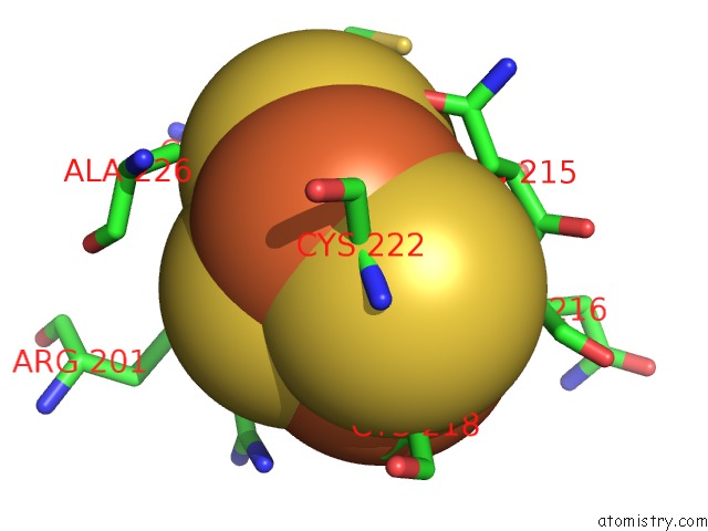
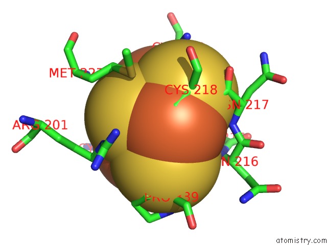
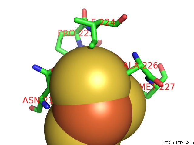
Iron binding site 1 out of 6 in 6a2u
Go back to
Iron binding site 1 out
of 6 in the Crystal Structure of Gamma-Alpha Subunit Complex From Burkholderia Cepacia Fad Glucose Dehydrogenase
Mono view
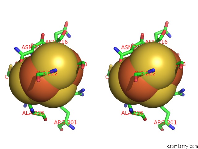
Stereo pair view
Mono view
Stereo pair view
A full contact list of Iron with other atoms in the Fe binding
site number 1 of Crystal Structure of Gamma-Alpha Subunit Complex From Burkholderia Cepacia Fad Glucose Dehydrogenase within 5.0Å range:
|
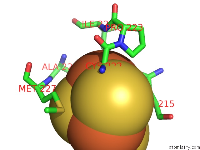
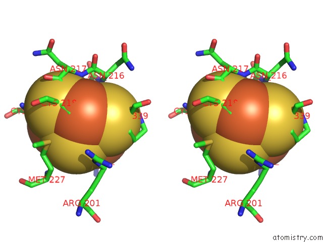
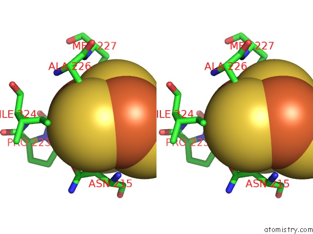
Iron binding site 2 out of 6 in 6a2u
Go back to
Iron binding site 2 out
of 6 in the Crystal Structure of Gamma-Alpha Subunit Complex From Burkholderia Cepacia Fad Glucose Dehydrogenase
Mono view
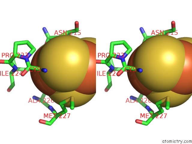
Stereo pair view
Mono view
Stereo pair view
A full contact list of Iron with other atoms in the Fe binding
site number 2 of Crystal Structure of Gamma-Alpha Subunit Complex From Burkholderia Cepacia Fad Glucose Dehydrogenase within 5.0Å range:
|
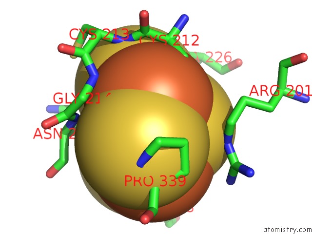
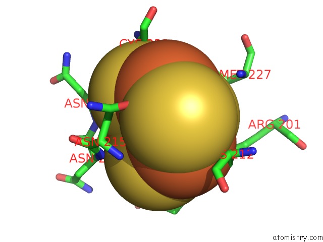
Iron binding site 3 out of 6 in 6a2u
Go back to
Iron binding site 3 out
of 6 in the Crystal Structure of Gamma-Alpha Subunit Complex From Burkholderia Cepacia Fad Glucose Dehydrogenase
Mono view
Stereo pair view
Mono view
Stereo pair view
A full contact list of Iron with other atoms in the Fe binding
site number 3 of Crystal Structure of Gamma-Alpha Subunit Complex From Burkholderia Cepacia Fad Glucose Dehydrogenase within 5.0Å range:
|
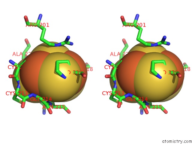
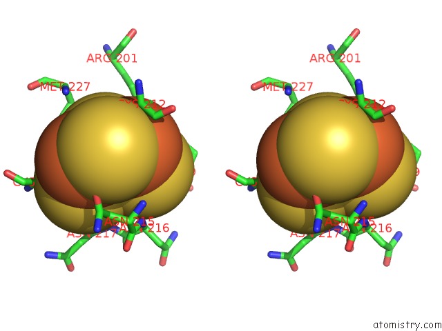
Iron binding site 4 out of 6 in 6a2u
Go back to
Iron binding site 4 out
of 6 in the Crystal Structure of Gamma-Alpha Subunit Complex From Burkholderia Cepacia Fad Glucose Dehydrogenase
Mono view
Stereo pair view
Mono view
Stereo pair view
A full contact list of Iron with other atoms in the Fe binding
site number 4 of Crystal Structure of Gamma-Alpha Subunit Complex From Burkholderia Cepacia Fad Glucose Dehydrogenase within 5.0Å range:
|
Iron binding site 5 out of 6 in 6a2u
Go back to
Iron binding site 5 out
of 6 in the Crystal Structure of Gamma-Alpha Subunit Complex From Burkholderia Cepacia Fad Glucose Dehydrogenase
Mono view
Stereo pair view
Mono view
Stereo pair view
A full contact list of Iron with other atoms in the Fe binding
site number 5 of Crystal Structure of Gamma-Alpha Subunit Complex From Burkholderia Cepacia Fad Glucose Dehydrogenase within 5.0Å range:
|
Iron binding site 6 out of 6 in 6a2u
Go back to
Iron binding site 6 out
of 6 in the Crystal Structure of Gamma-Alpha Subunit Complex From Burkholderia Cepacia Fad Glucose Dehydrogenase
Mono view
Stereo pair view
Mono view
Stereo pair view
A full contact list of Iron with other atoms in the Fe binding
site number 6 of Crystal Structure of Gamma-Alpha Subunit Complex From Burkholderia Cepacia Fad Glucose Dehydrogenase within 5.0Å range:
|
Reference:
H.Yoshida,
K.Kojima,
M.Shiota,
K.Yoshimatsu,
T.Yamazaki,
S.Ferri,
W.Tsugawa,
S.Kamitori,
K.Sode.
X-Ray Structure of the Direct Electron Transfer-Type Fad Glucose Dehydrogenase Catalytic Subunit Complexed with A Hitchhiker Protein. Acta Crystallogr D Struct V. 75 841 2019BIOL.
ISSN: ISSN 2059-7983
PubMed: 31478907
DOI: 10.1107/S2059798319010878
Page generated: Wed Aug 6 03:40:11 2025
ISSN: ISSN 2059-7983
PubMed: 31478907
DOI: 10.1107/S2059798319010878
Last articles
Mg in 1IR1Mg in 1IR3
Mg in 1IQC
Mg in 1IBL
Mg in 1IPW
Mg in 1IQ8
Mg in 1IPP
Mg in 1IOW
Mg in 1IBM
Mg in 1IOV