Iron »
PDB 6ajo-6ayc »
6ajp »
Iron in PDB 6ajp: Complex Form of Uracil Dna Glycosylase X and Deoxyuridine Monophosphate.
Protein crystallography data
The structure of Complex Form of Uracil Dna Glycosylase X and Deoxyuridine Monophosphate., PDB code: 6ajp
was solved by
W.C.Ahn,
S.Aroli,
U.Varshney,
E.J.Woo,
with X-Ray Crystallography technique. A brief refinement statistics is given in the table below:
| Resolution Low / High (Å) | 18.88 / 1.33 |
| Space group | P 1 21 1 |
| Cell size a, b, c (Å), α, β, γ (°) | 36.436, 51.209, 54.533, 90.00, 104.98, 90.00 |
| R / Rfree (%) | 16.3 / 18.7 |
Iron Binding Sites:
The binding sites of Iron atom in the Complex Form of Uracil Dna Glycosylase X and Deoxyuridine Monophosphate.
(pdb code 6ajp). This binding sites where shown within
5.0 Angstroms radius around Iron atom.
In total 4 binding sites of Iron where determined in the Complex Form of Uracil Dna Glycosylase X and Deoxyuridine Monophosphate., PDB code: 6ajp:
Jump to Iron binding site number: 1; 2; 3; 4;
In total 4 binding sites of Iron where determined in the Complex Form of Uracil Dna Glycosylase X and Deoxyuridine Monophosphate., PDB code: 6ajp:
Jump to Iron binding site number: 1; 2; 3; 4;
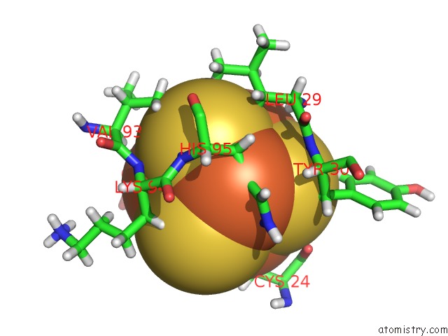
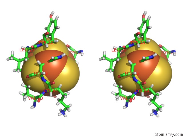
Iron binding site 1 out of 4 in 6ajp
Go back to
Iron binding site 1 out
of 4 in the Complex Form of Uracil Dna Glycosylase X and Deoxyuridine Monophosphate.
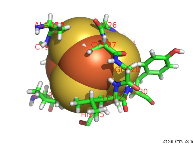
Mono view
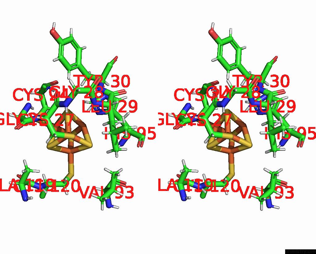
Stereo pair view
Mono view
Stereo pair view
A full contact list of Iron with other atoms in the Fe binding
site number 1 of Complex Form of Uracil Dna Glycosylase X and Deoxyuridine Monophosphate. within 5.0Å range:
|
Iron binding site 2 out of 4 in 6ajp
Go back to
Iron binding site 2 out
of 4 in the Complex Form of Uracil Dna Glycosylase X and Deoxyuridine Monophosphate.
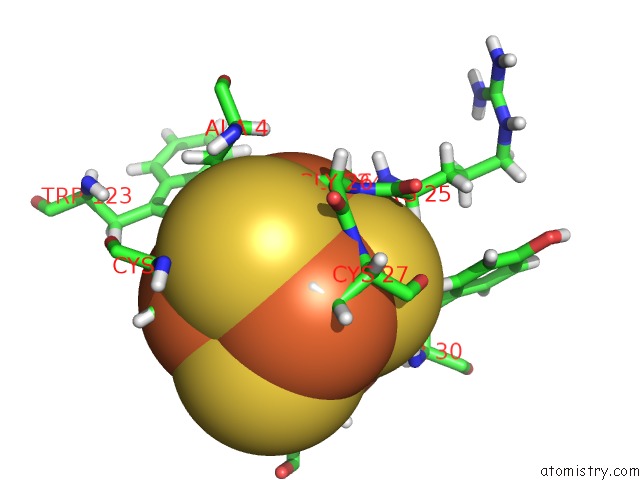
Mono view
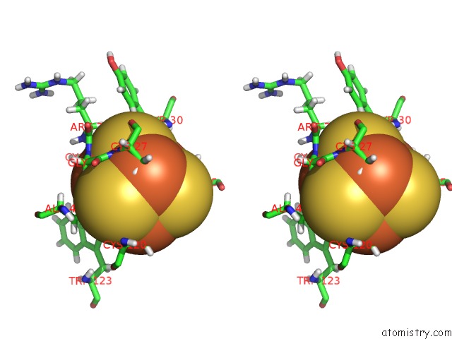
Stereo pair view
Mono view
Stereo pair view
A full contact list of Iron with other atoms in the Fe binding
site number 2 of Complex Form of Uracil Dna Glycosylase X and Deoxyuridine Monophosphate. within 5.0Å range:
|
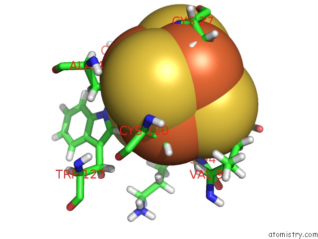
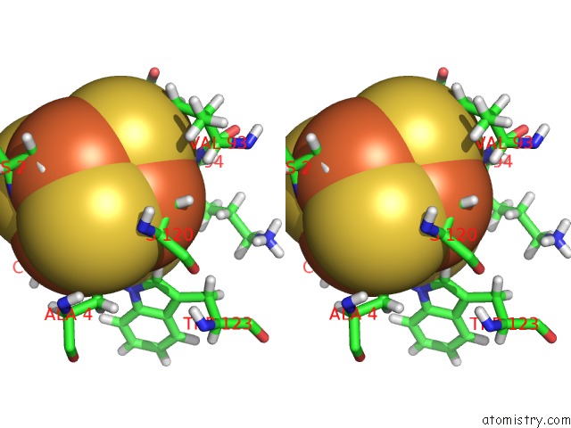
Iron binding site 3 out of 4 in 6ajp
Go back to
Iron binding site 3 out
of 4 in the Complex Form of Uracil Dna Glycosylase X and Deoxyuridine Monophosphate.
Mono view
Stereo pair view
Mono view
Stereo pair view
A full contact list of Iron with other atoms in the Fe binding
site number 3 of Complex Form of Uracil Dna Glycosylase X and Deoxyuridine Monophosphate. within 5.0Å range:
|
Iron binding site 4 out of 4 in 6ajp
Go back to
Iron binding site 4 out
of 4 in the Complex Form of Uracil Dna Glycosylase X and Deoxyuridine Monophosphate.
Mono view
Stereo pair view
Mono view
Stereo pair view
A full contact list of Iron with other atoms in the Fe binding
site number 4 of Complex Form of Uracil Dna Glycosylase X and Deoxyuridine Monophosphate. within 5.0Å range:
|
Reference:
W.C.Ahn,
S.Aroli,
J.H.Kim,
J.H.Moon,
G.S.Lee,
M.H.Lee,
P.B.Sang,
B.H.Oh,
U.Varshney,
E.J.Woo.
Covalent Binding of Uracil Dna Glycosylase Udgx to Abasic Dna Upon Uracil Excision. Nat.Chem.Biol. V. 15 607 2019.
ISSN: ESSN 1552-4469
PubMed: 31101917
DOI: 10.1038/S41589-019-0289-3
Page generated: Wed Aug 6 03:49:03 2025
ISSN: ESSN 1552-4469
PubMed: 31101917
DOI: 10.1038/S41589-019-0289-3
Last articles
Mn in 8F4CMn in 8EZ5
Mn in 8ECY
Mn in 8EQM
Mn in 8EIP
Mn in 8EPZ
Mn in 8EIN
Mn in 8E8H
Mn in 8EDZ
Mn in 8E8G