Iron »
PDB 6azu-6bpt »
6bde »
Iron in PDB 6bde: Crystal Structure of Fe(II) Unliganded H-Nox Protein Mutant A71G From K. Algicida
Protein crystallography data
The structure of Crystal Structure of Fe(II) Unliganded H-Nox Protein Mutant A71G From K. Algicida, PDB code: 6bde
was solved by
J.J.Bruegger,
C.W.Hespen,
M.A.Marletta,
with X-Ray Crystallography technique. A brief refinement statistics is given in the table below:
| Resolution Low / High (Å) | 39.79 / 1.64 |
| Space group | P 21 21 21 |
| Cell size a, b, c (Å), α, β, γ (°) | 48.794, 56.730, 68.753, 90.00, 90.00, 90.00 |
| R / Rfree (%) | 21 / 23.6 |
Iron Binding Sites:
The binding sites of Iron atom in the Crystal Structure of Fe(II) Unliganded H-Nox Protein Mutant A71G From K. Algicida
(pdb code 6bde). This binding sites where shown within
5.0 Angstroms radius around Iron atom.
In total only one binding site of Iron was determined in the Crystal Structure of Fe(II) Unliganded H-Nox Protein Mutant A71G From K. Algicida, PDB code: 6bde:
In total only one binding site of Iron was determined in the Crystal Structure of Fe(II) Unliganded H-Nox Protein Mutant A71G From K. Algicida, PDB code: 6bde:
Iron binding site 1 out of 1 in 6bde
Go back to
Iron binding site 1 out
of 1 in the Crystal Structure of Fe(II) Unliganded H-Nox Protein Mutant A71G From K. Algicida
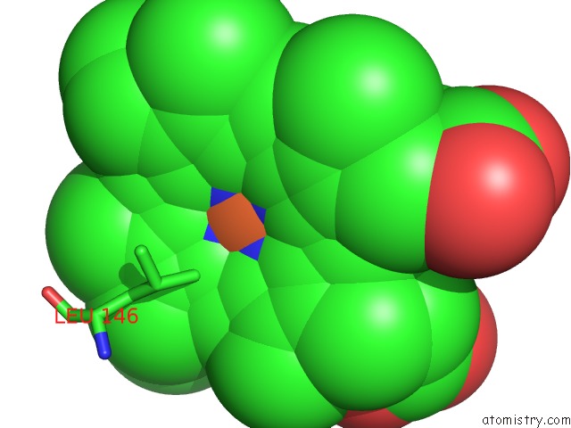
Mono view
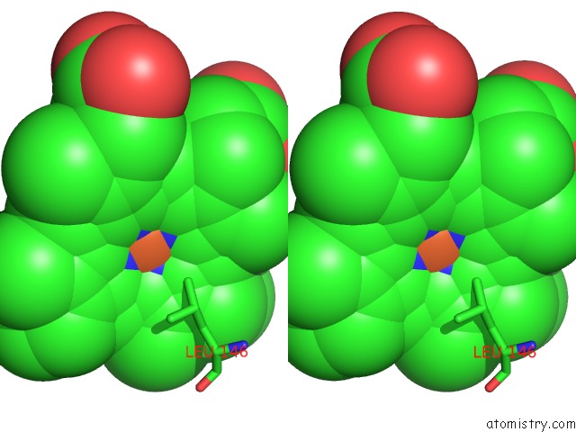
Stereo pair view
Mono view
Stereo pair view
A full contact list of Iron with other atoms in the Fe binding
site number 1 of Crystal Structure of Fe(II) Unliganded H-Nox Protein Mutant A71G From K. Algicida within 5.0Å range:
|
Reference:
C.W.Hespen,
J.J.Bruegger,
Y.Guo,
M.A.Marletta.
Native Alanine Substitution in the Glycine Hinge Modulates Conformational Flexibility of Heme Nitric Oxide/Oxygen (H-Nox) Sensing Proteins. Acs Chem. Biol. V. 13 1631 2018.
ISSN: ESSN 1554-8937
PubMed: 29757599
DOI: 10.1021/ACSCHEMBIO.8B00248
Page generated: Wed Aug 6 04:07:26 2025
ISSN: ESSN 1554-8937
PubMed: 29757599
DOI: 10.1021/ACSCHEMBIO.8B00248
Last articles
Na in 5TIBNa in 5TEQ
Na in 5TFM
Na in 5TEJ
Na in 5TFL
Na in 5TEK
Na in 5TE1
Na in 5TDR
Na in 5TEE
Na in 5TDW