Iron »
PDB 6azu-6bpt »
6bmt »
Iron in PDB 6bmt: Crystal Structure of A Recombinant Form of Human Myeloperoxidase Bound to An Inhibitor From Staphylococcus Delphini
Enzymatic activity of Crystal Structure of A Recombinant Form of Human Myeloperoxidase Bound to An Inhibitor From Staphylococcus Delphini
All present enzymatic activity of Crystal Structure of A Recombinant Form of Human Myeloperoxidase Bound to An Inhibitor From Staphylococcus Delphini:
1.11.2.2;
1.11.2.2;
Protein crystallography data
The structure of Crystal Structure of A Recombinant Form of Human Myeloperoxidase Bound to An Inhibitor From Staphylococcus Delphini, PDB code: 6bmt
was solved by
N.T.Ploscariu,
B.V.Geisbrecht,
with X-Ray Crystallography technique. A brief refinement statistics is given in the table below:
| Resolution Low / High (Å) | 45.34 / 2.40 |
| Space group | P 21 21 21 |
| Cell size a, b, c (Å), α, β, γ (°) | 84.626, 90.674, 125.661, 90.00, 90.00, 90.00 |
| R / Rfree (%) | 18.5 / 21.8 |
Other elements in 6bmt:
The structure of Crystal Structure of A Recombinant Form of Human Myeloperoxidase Bound to An Inhibitor From Staphylococcus Delphini also contains other interesting chemical elements:
| Chlorine | (Cl) | 1 atom |
| Calcium | (Ca) | 1 atom |
Iron Binding Sites:
The binding sites of Iron atom in the Crystal Structure of A Recombinant Form of Human Myeloperoxidase Bound to An Inhibitor From Staphylococcus Delphini
(pdb code 6bmt). This binding sites where shown within
5.0 Angstroms radius around Iron atom.
In total only one binding site of Iron was determined in the Crystal Structure of A Recombinant Form of Human Myeloperoxidase Bound to An Inhibitor From Staphylococcus Delphini, PDB code: 6bmt:
In total only one binding site of Iron was determined in the Crystal Structure of A Recombinant Form of Human Myeloperoxidase Bound to An Inhibitor From Staphylococcus Delphini, PDB code: 6bmt:
Iron binding site 1 out of 1 in 6bmt
Go back to
Iron binding site 1 out
of 1 in the Crystal Structure of A Recombinant Form of Human Myeloperoxidase Bound to An Inhibitor From Staphylococcus Delphini
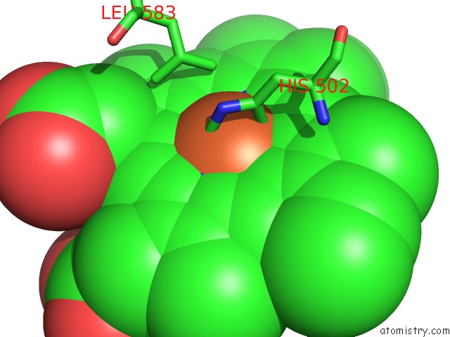
Mono view
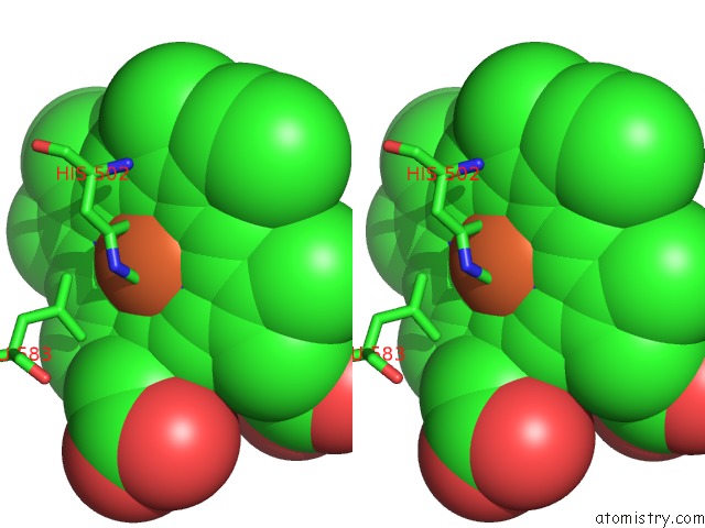
Stereo pair view
Mono view
Stereo pair view
A full contact list of Iron with other atoms in the Fe binding
site number 1 of Crystal Structure of A Recombinant Form of Human Myeloperoxidase Bound to An Inhibitor From Staphylococcus Delphini within 5.0Å range:
|
Reference:
N.T.Ploscariu,
N.W.M.De Jong,
K.P.M.Van Kessel,
J.A.G.Van Strijp,
B.V.Geisbrecht.
Identification and Structural Characterization of A Novel Myeloperoxidase Inhibitor From Staphylococcus Delphini. Arch. Biochem. Biophys. V. 645 1 2018.
ISSN: ESSN 1096-0384
PubMed: 29524428
DOI: 10.1016/J.ABB.2018.03.007
Page generated: Wed Aug 6 04:09:36 2025
ISSN: ESSN 1096-0384
PubMed: 29524428
DOI: 10.1016/J.ABB.2018.03.007
Last articles
Fe in 7BLXFe in 7BKD
Fe in 7BI7
Fe in 7BHC
Fe in 7BIU
Fe in 7BI1
Fe in 7B9A
Fe in 7BGI
Fe in 7BHB
Fe in 7B97