Iron »
PDB 6e45-6f0a »
6e4v »
Iron in PDB 6e4v: The Crystal Structure of Fhue From E. Coli in Complex with Its Substrate Coprogen
Protein crystallography data
The structure of The Crystal Structure of Fhue From E. Coli in Complex with Its Substrate Coprogen, PDB code: 6e4v
was solved by
R.Grinter,
T.Lithgow,
with X-Ray Crystallography technique. A brief refinement statistics is given in the table below:
| Resolution Low / High (Å) | 47.57 / 2.00 |
| Space group | P 21 21 21 |
| Cell size a, b, c (Å), α, β, γ (°) | 68.190, 103.860, 118.620, 90.00, 90.00, 90.00 |
| R / Rfree (%) | 20.6 / 23.5 |
Iron Binding Sites:
The binding sites of Iron atom in the The Crystal Structure of Fhue From E. Coli in Complex with Its Substrate Coprogen
(pdb code 6e4v). This binding sites where shown within
5.0 Angstroms radius around Iron atom.
In total only one binding site of Iron was determined in the The Crystal Structure of Fhue From E. Coli in Complex with Its Substrate Coprogen, PDB code: 6e4v:
In total only one binding site of Iron was determined in the The Crystal Structure of Fhue From E. Coli in Complex with Its Substrate Coprogen, PDB code: 6e4v:
Iron binding site 1 out of 1 in 6e4v
Go back to
Iron binding site 1 out
of 1 in the The Crystal Structure of Fhue From E. Coli in Complex with Its Substrate Coprogen
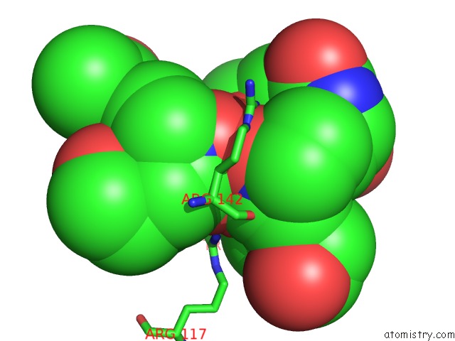
Mono view
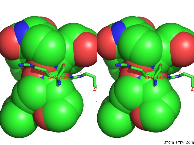
Stereo pair view
Mono view
Stereo pair view
A full contact list of Iron with other atoms in the Fe binding
site number 1 of The Crystal Structure of Fhue From E. Coli in Complex with Its Substrate Coprogen within 5.0Å range:
|
Reference:
R.Grinter,
T.Lithgow.
Determination of the Molecular Basis For Coprogen Import By Gram-Negative Bacteria. Iucrj V. 6 401 2019.
ISSN: ESSN 2052-2525
PubMed: 31098021
DOI: 10.1107/S2052252519002926
Page generated: Tue Aug 6 17:18:14 2024
ISSN: ESSN 2052-2525
PubMed: 31098021
DOI: 10.1107/S2052252519002926
Last articles
Fe in 2YXOFe in 2YRS
Fe in 2YXC
Fe in 2YNM
Fe in 2YVJ
Fe in 2YP1
Fe in 2YU2
Fe in 2YU1
Fe in 2YQB
Fe in 2YOO