Iron »
PDB 6fmo-6g5t »
6frm »
Iron in PDB 6frm: Crystal Structure of Coenzyme F420H2 Oxidase (Fpra) Co-Crystallized with 10 Mm Tb-XO4
Protein crystallography data
The structure of Crystal Structure of Coenzyme F420H2 Oxidase (Fpra) Co-Crystallized with 10 Mm Tb-XO4, PDB code: 6frm
was solved by
S.Engilberge,
F.Riobe,
T.Wagner,
S.Di Pietro,
S.Shima,
E.Girard,
E.Dumont,
O.Maury,
with X-Ray Crystallography technique. A brief refinement statistics is given in the table below:
| Resolution Low / High (Å) | 48.69 / 2.20 |
| Space group | P 1 21 1 |
| Cell size a, b, c (Å), α, β, γ (°) | 84.170, 147.875, 146.059, 90.00, 90.40, 90.00 |
| R / Rfree (%) | 17.7 / 20.9 |
Other elements in 6frm:
The structure of Crystal Structure of Coenzyme F420H2 Oxidase (Fpra) Co-Crystallized with 10 Mm Tb-XO4 also contains other interesting chemical elements:
| Terbium | (Tb) | 16 atoms |
| Chlorine | (Cl) | 17 atoms |
Iron Binding Sites:
The binding sites of Iron atom in the Crystal Structure of Coenzyme F420H2 Oxidase (Fpra) Co-Crystallized with 10 Mm Tb-XO4
(pdb code 6frm). This binding sites where shown within
5.0 Angstroms radius around Iron atom.
In total 4 binding sites of Iron where determined in the Crystal Structure of Coenzyme F420H2 Oxidase (Fpra) Co-Crystallized with 10 Mm Tb-XO4, PDB code: 6frm:
Jump to Iron binding site number: 1; 2; 3; 4;
In total 4 binding sites of Iron where determined in the Crystal Structure of Coenzyme F420H2 Oxidase (Fpra) Co-Crystallized with 10 Mm Tb-XO4, PDB code: 6frm:
Jump to Iron binding site number: 1; 2; 3; 4;
Iron binding site 1 out of 4 in 6frm
Go back to
Iron binding site 1 out
of 4 in the Crystal Structure of Coenzyme F420H2 Oxidase (Fpra) Co-Crystallized with 10 Mm Tb-XO4
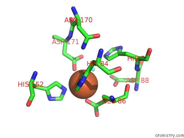
Mono view
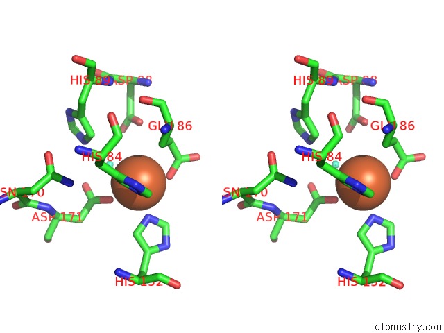
Stereo pair view
Mono view
Stereo pair view
A full contact list of Iron with other atoms in the Fe binding
site number 1 of Crystal Structure of Coenzyme F420H2 Oxidase (Fpra) Co-Crystallized with 10 Mm Tb-XO4 within 5.0Å range:
|
Iron binding site 2 out of 4 in 6frm
Go back to
Iron binding site 2 out
of 4 in the Crystal Structure of Coenzyme F420H2 Oxidase (Fpra) Co-Crystallized with 10 Mm Tb-XO4

Mono view

Stereo pair view
Mono view
Stereo pair view
A full contact list of Iron with other atoms in the Fe binding
site number 2 of Crystal Structure of Coenzyme F420H2 Oxidase (Fpra) Co-Crystallized with 10 Mm Tb-XO4 within 5.0Å range:
|
Iron binding site 3 out of 4 in 6frm
Go back to
Iron binding site 3 out
of 4 in the Crystal Structure of Coenzyme F420H2 Oxidase (Fpra) Co-Crystallized with 10 Mm Tb-XO4

Mono view

Stereo pair view
Mono view
Stereo pair view
A full contact list of Iron with other atoms in the Fe binding
site number 3 of Crystal Structure of Coenzyme F420H2 Oxidase (Fpra) Co-Crystallized with 10 Mm Tb-XO4 within 5.0Å range:
|
Iron binding site 4 out of 4 in 6frm
Go back to
Iron binding site 4 out
of 4 in the Crystal Structure of Coenzyme F420H2 Oxidase (Fpra) Co-Crystallized with 10 Mm Tb-XO4

Mono view

Stereo pair view
Mono view
Stereo pair view
A full contact list of Iron with other atoms in the Fe binding
site number 4 of Crystal Structure of Coenzyme F420H2 Oxidase (Fpra) Co-Crystallized with 10 Mm Tb-XO4 within 5.0Å range:
|
Reference:
S.Engilberge,
F.Riobe,
T.Wagner,
S.Di Pietro,
C.Breyton,
B.Franzetti,
S.Shima,
E.Girard,
E.Dumont,
O.Maury.
Unveiling the Binding Modes of the Crystallophore, A Terbium-Based Nucleating and Phasing Molecular Agent For Protein Crystallography. Chemistry V. 24 9739 2018.
ISSN: ISSN 1521-3765
PubMed: 29806881
DOI: 10.1002/CHEM.201802172
Page generated: Wed Aug 6 06:16:55 2025
ISSN: ISSN 1521-3765
PubMed: 29806881
DOI: 10.1002/CHEM.201802172
Last articles
Mn in 9LJUMn in 9LJW
Mn in 9LJS
Mn in 9LJR
Mn in 9LJT
Mn in 9LJV
Mg in 9UA2
Mg in 9R96
Mg in 9VM1
Mg in 9P01