Iron »
PDB 6g71-6gl6 »
6gii »
Iron in PDB 6gii: The Crystal Structure of Tepidiphilus Thermophilus P450 Heme Domain
Protein crystallography data
The structure of The Crystal Structure of Tepidiphilus Thermophilus P450 Heme Domain, PDB code: 6gii
was solved by
C.W.Levy,
with X-Ray Crystallography technique. A brief refinement statistics is given in the table below:
| Resolution Low / High (Å) | 43.32 / 1.90 |
| Space group | P 21 21 21 |
| Cell size a, b, c (Å), α, β, γ (°) | 53.292, 74.384, 105.013, 90.00, 90.00, 90.00 |
| R / Rfree (%) | 17.9 / 22.8 |
Iron Binding Sites:
The binding sites of Iron atom in the The Crystal Structure of Tepidiphilus Thermophilus P450 Heme Domain
(pdb code 6gii). This binding sites where shown within
5.0 Angstroms radius around Iron atom.
In total only one binding site of Iron was determined in the The Crystal Structure of Tepidiphilus Thermophilus P450 Heme Domain, PDB code: 6gii:
In total only one binding site of Iron was determined in the The Crystal Structure of Tepidiphilus Thermophilus P450 Heme Domain, PDB code: 6gii:
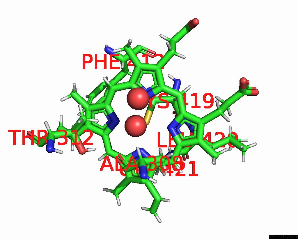
Iron binding site 1 out of 1 in 6gii
Go back to
Iron binding site 1 out
of 1 in the The Crystal Structure of Tepidiphilus Thermophilus P450 Heme Domain
Mono view

Stereo pair view
Mono view
Stereo pair view
A full contact list of Iron with other atoms in the Fe binding
site number 1 of The Crystal Structure of Tepidiphilus Thermophilus P450 Heme Domain within 5.0Å range:
|
Reference:
M.Tavanti,
J.L.Porter,
C.W.Levy,
J.R.Gomez Castellanos,
S.L.Flitsch,
N.J.Turner.
The Crystal Structure of P450-Tt Heme-Domain Provides the First Structural Insights Into the Versatile Class VII P450S. Biochem. Biophys. Res. V. 501 846 2018COMMUN..
ISSN: ESSN 1090-2104
PubMed: 29738765
DOI: 10.1016/J.BBRC.2018.05.014
Page generated: Wed Aug 6 06:52:37 2025
ISSN: ESSN 1090-2104
PubMed: 29738765
DOI: 10.1016/J.BBRC.2018.05.014
Last articles
Zn in 9QM9Zn in 9S44
Zn in 9OFE
Zn in 9OFC
Zn in 9OFD
Zn in 9OF1
Zn in 9OFB
Zn in 9N0J
Zn in 9M5X
Zn in 9LGI