Iron »
PDB 6gly-6haw »
6gmf »
Iron in PDB 6gmf: Structure of Cytochrome P450 CYP109Q5 From Chondromyces Apiculatus
Protein crystallography data
The structure of Structure of Cytochrome P450 CYP109Q5 From Chondromyces Apiculatus, PDB code: 6gmf
was solved by
G.Grogan,
P.Dubiel,
M.Sharma,
J.Klenk,
B.Hauer,
with X-Ray Crystallography technique. A brief refinement statistics is given in the table below:
| Resolution Low / High (Å) | 44.71 / 1.55 |
| Space group | P 1 21 1 |
| Cell size a, b, c (Å), α, β, γ (°) | 44.690, 55.020, 74.800, 90.00, 91.67, 90.00 |
| R / Rfree (%) | 18.5 / 22 |
Iron Binding Sites:
The binding sites of Iron atom in the Structure of Cytochrome P450 CYP109Q5 From Chondromyces Apiculatus
(pdb code 6gmf). This binding sites where shown within
5.0 Angstroms radius around Iron atom.
In total only one binding site of Iron was determined in the Structure of Cytochrome P450 CYP109Q5 From Chondromyces Apiculatus, PDB code: 6gmf:
In total only one binding site of Iron was determined in the Structure of Cytochrome P450 CYP109Q5 From Chondromyces Apiculatus, PDB code: 6gmf:
Iron binding site 1 out of 1 in 6gmf
Go back to
Iron binding site 1 out
of 1 in the Structure of Cytochrome P450 CYP109Q5 From Chondromyces Apiculatus
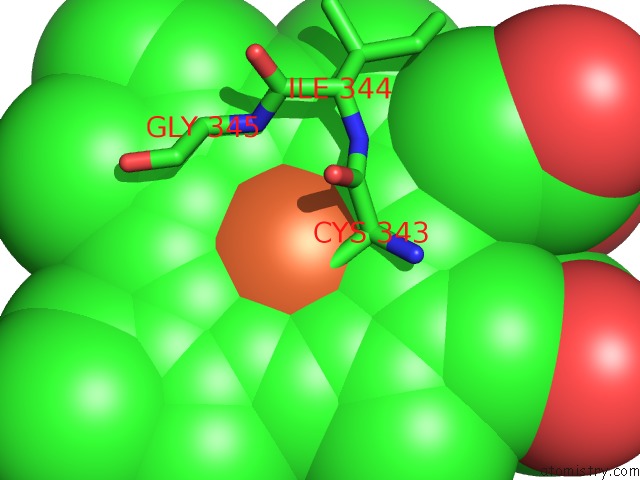
Mono view
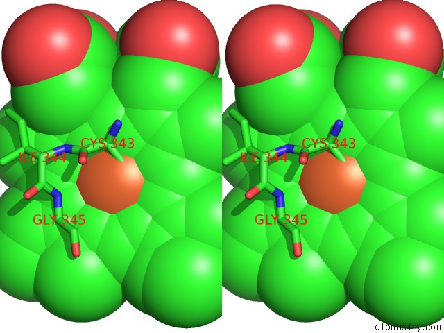
Stereo pair view
Mono view
Stereo pair view
A full contact list of Iron with other atoms in the Fe binding
site number 1 of Structure of Cytochrome P450 CYP109Q5 From Chondromyces Apiculatus within 5.0Å range:
|
Reference:
J.M.Klenk,
P.Dubiel,
M.Sharma,
G.Grogan,
B.Hauer.
Characterization and Structure-Guided Engineering of the Novel Versatile Terpene Monooxygenase CYP109Q5 From Chondromyces Apiculatus DSM436. Microb Biotechnol V. 12 377 2019.
ISSN: ISSN 1751-7915
PubMed: 30592153
DOI: 10.1111/1751-7915.13354
Page generated: Wed Aug 6 07:25:28 2025
ISSN: ISSN 1751-7915
PubMed: 30592153
DOI: 10.1111/1751-7915.13354
Last articles
Na in 1KY8Na in 1L0I
Na in 1KVR
Na in 1KVS
Na in 1KVT
Na in 1KTW
Na in 1KVQ
Na in 1KSU
Na in 1KR3
Na in 1KSS