Iron »
PDB 6gly-6haw »
6gpe »
Iron in PDB 6gpe: Crystal Structure of the Csid Glutarate Hydroxylase
Protein crystallography data
The structure of Crystal Structure of the Csid Glutarate Hydroxylase, PDB code: 6gpe
was solved by
R.M.Williams,
O.Mayans,
J.S.Hartig,
with X-Ray Crystallography technique. A brief refinement statistics is given in the table below:
| Resolution Low / High (Å) | 42.51 / 2.20 |
| Space group | P 4 21 2 |
| Cell size a, b, c (Å), α, β, γ (°) | 121.200, 121.200, 137.000, 90.00, 90.00, 90.00 |
| R / Rfree (%) | 17.3 / 19.8 |
Iron Binding Sites:
The binding sites of Iron atom in the Crystal Structure of the Csid Glutarate Hydroxylase
(pdb code 6gpe). This binding sites where shown within
5.0 Angstroms radius around Iron atom.
In total 2 binding sites of Iron where determined in the Crystal Structure of the Csid Glutarate Hydroxylase, PDB code: 6gpe:
Jump to Iron binding site number: 1; 2;
In total 2 binding sites of Iron where determined in the Crystal Structure of the Csid Glutarate Hydroxylase, PDB code: 6gpe:
Jump to Iron binding site number: 1; 2;
Iron binding site 1 out of 2 in 6gpe
Go back to
Iron binding site 1 out
of 2 in the Crystal Structure of the Csid Glutarate Hydroxylase
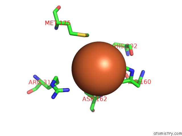
Mono view
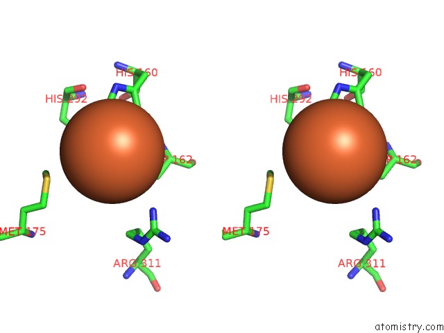
Stereo pair view
Mono view
Stereo pair view
A full contact list of Iron with other atoms in the Fe binding
site number 1 of Crystal Structure of the Csid Glutarate Hydroxylase within 5.0Å range:
|
Iron binding site 2 out of 2 in 6gpe
Go back to
Iron binding site 2 out
of 2 in the Crystal Structure of the Csid Glutarate Hydroxylase
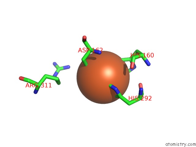
Mono view
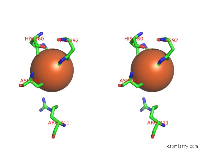
Stereo pair view
Mono view
Stereo pair view
A full contact list of Iron with other atoms in the Fe binding
site number 2 of Crystal Structure of the Csid Glutarate Hydroxylase within 5.0Å range:
|
Reference:
S.Knorr,
M.Sinn,
D.Galetskiy,
R.M.Williams,
C.Wang,
N.Muller,
O.Mayans,
D.Schleheck,
J.S.Hartig.
Widespread Bacterial Lysine Degradation Proceeding Via Glutarate and L-2-Hydroxyglutarate. Nat Commun V. 9 5071 2018.
ISSN: ESSN 2041-1723
PubMed: 30498244
DOI: 10.1038/S41467-018-07563-6
Page generated: Wed Aug 6 07:25:53 2025
ISSN: ESSN 2041-1723
PubMed: 30498244
DOI: 10.1038/S41467-018-07563-6
Last articles
Mn in 8ZWDMn in 8ZX9
Mn in 8ZX8
Mn in 8ZX2
Mn in 8ZX3
Mn in 8ZWY
Mn in 8ZWW
Mn in 8ZRL
Mn in 8ZWR
Mn in 8ZWP