Iron »
PDB 6htk-6i93 »
6i0r »
Iron in PDB 6i0r: Structure of Quinolinate Synthase in Complex with 5-Mercaptopyridine- 2,3-Dicarboxylic Acid
Enzymatic activity of Structure of Quinolinate Synthase in Complex with 5-Mercaptopyridine- 2,3-Dicarboxylic Acid
All present enzymatic activity of Structure of Quinolinate Synthase in Complex with 5-Mercaptopyridine- 2,3-Dicarboxylic Acid:
2.5.1.72;
2.5.1.72;
Protein crystallography data
The structure of Structure of Quinolinate Synthase in Complex with 5-Mercaptopyridine- 2,3-Dicarboxylic Acid, PDB code: 6i0r
was solved by
A.Volbeda,
J.C.Fontecilla-Camps,
with X-Ray Crystallography technique. A brief refinement statistics is given in the table below:
| Resolution Low / High (Å) | 37.16 / 2.10 |
| Space group | P 1 21 1 |
| Cell size a, b, c (Å), α, β, γ (°) | 54.900, 48.500, 60.500, 90.00, 107.10, 90.00 |
| R / Rfree (%) | 19.8 / 23.9 |
Iron Binding Sites:
The binding sites of Iron atom in the Structure of Quinolinate Synthase in Complex with 5-Mercaptopyridine- 2,3-Dicarboxylic Acid
(pdb code 6i0r). This binding sites where shown within
5.0 Angstroms radius around Iron atom.
In total 4 binding sites of Iron where determined in the Structure of Quinolinate Synthase in Complex with 5-Mercaptopyridine- 2,3-Dicarboxylic Acid, PDB code: 6i0r:
Jump to Iron binding site number: 1; 2; 3; 4;
In total 4 binding sites of Iron where determined in the Structure of Quinolinate Synthase in Complex with 5-Mercaptopyridine- 2,3-Dicarboxylic Acid, PDB code: 6i0r:
Jump to Iron binding site number: 1; 2; 3; 4;
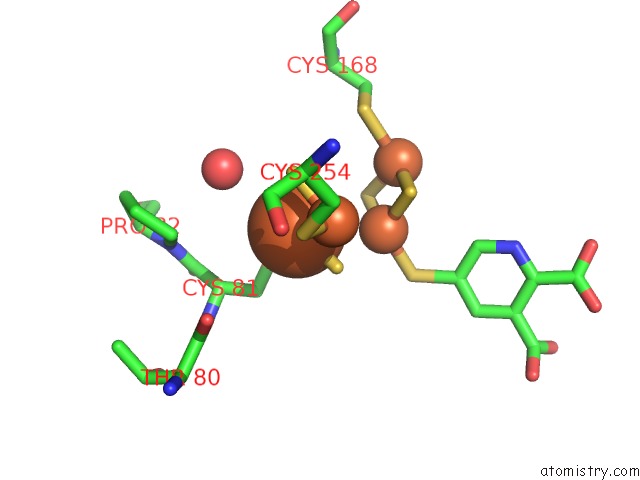
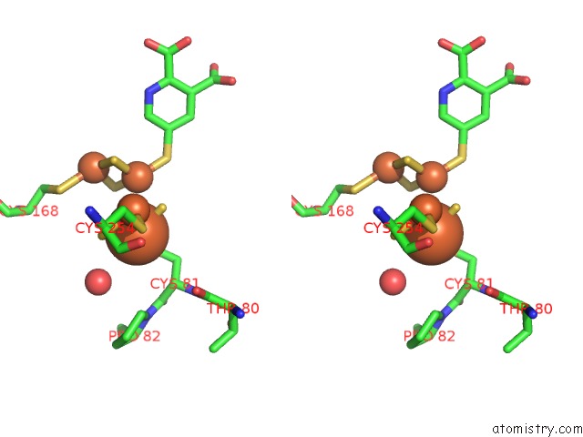
Iron binding site 1 out of 4 in 6i0r
Go back to
Iron binding site 1 out
of 4 in the Structure of Quinolinate Synthase in Complex with 5-Mercaptopyridine- 2,3-Dicarboxylic Acid
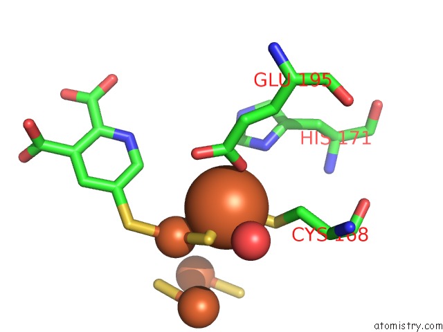
Mono view
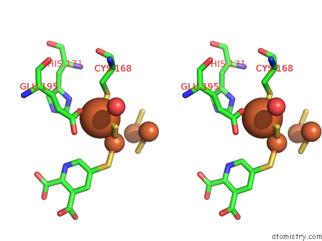
Stereo pair view
Mono view
Stereo pair view
A full contact list of Iron with other atoms in the Fe binding
site number 1 of Structure of Quinolinate Synthase in Complex with 5-Mercaptopyridine- 2,3-Dicarboxylic Acid within 5.0Å range:
|
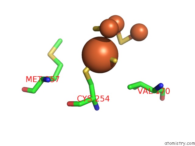
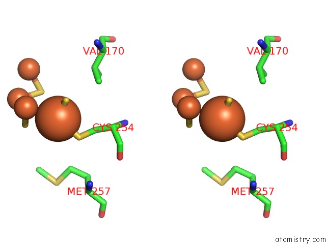
Iron binding site 2 out of 4 in 6i0r
Go back to
Iron binding site 2 out
of 4 in the Structure of Quinolinate Synthase in Complex with 5-Mercaptopyridine- 2,3-Dicarboxylic Acid
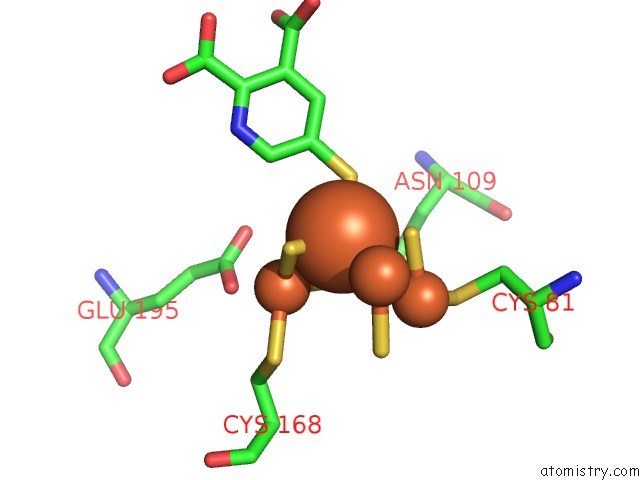
Mono view
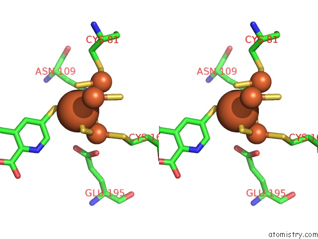
Stereo pair view
Mono view
Stereo pair view
A full contact list of Iron with other atoms in the Fe binding
site number 2 of Structure of Quinolinate Synthase in Complex with 5-Mercaptopyridine- 2,3-Dicarboxylic Acid within 5.0Å range:
|
Iron binding site 3 out of 4 in 6i0r
Go back to
Iron binding site 3 out
of 4 in the Structure of Quinolinate Synthase in Complex with 5-Mercaptopyridine- 2,3-Dicarboxylic Acid
Mono view
Stereo pair view
Mono view
Stereo pair view
A full contact list of Iron with other atoms in the Fe binding
site number 3 of Structure of Quinolinate Synthase in Complex with 5-Mercaptopyridine- 2,3-Dicarboxylic Acid within 5.0Å range:
|
Iron binding site 4 out of 4 in 6i0r
Go back to
Iron binding site 4 out
of 4 in the Structure of Quinolinate Synthase in Complex with 5-Mercaptopyridine- 2,3-Dicarboxylic Acid
Mono view
Stereo pair view
Mono view
Stereo pair view
A full contact list of Iron with other atoms in the Fe binding
site number 4 of Structure of Quinolinate Synthase in Complex with 5-Mercaptopyridine- 2,3-Dicarboxylic Acid within 5.0Å range:
|
Reference:
J.Saez Cabodevilla,
A.Volbeda,
O.Hamelin,
J.M.Latour,
O.Gigarel,
M.Clemancey,
C.Darnault,
D.Reichmann,
P.Amara,
J.C.Fontecilla-Camps,
S.Ollagnier De Choudens.
Design of Specific Inhibitors of Quinolinate Synthase Based on [4FE-4S] Cluster Coordination. Chem.Commun.(Camb.) V. 55 3725 2019.
ISSN: ESSN 1364-548X
PubMed: 30855610
DOI: 10.1039/C8CC09023H
Page generated: Tue Aug 6 22:00:39 2024
ISSN: ESSN 1364-548X
PubMed: 30855610
DOI: 10.1039/C8CC09023H
Last articles
Fe in 2YXOFe in 2YRS
Fe in 2YXC
Fe in 2YNM
Fe in 2YVJ
Fe in 2YP1
Fe in 2YU2
Fe in 2YU1
Fe in 2YQB
Fe in 2YOO