Iron »
PDB 6htk-6i93 »
6i7c »
Iron in PDB 6i7c: Dye Type Peroxidase Aa From Streptomyces Lividans: Imidazole Complex
Protein crystallography data
The structure of Dye Type Peroxidase Aa From Streptomyces Lividans: Imidazole Complex, PDB code: 6i7c
was solved by
T.Moreno-Chicano,
A.E.Ebrahim,
J.A.R.Worrall,
R.W.Strange,
D.Axford,
D.A.Sherrell,
H.Sugimoto,
K.Tono,
S.Owada,
H.Duyvesteyn,
with X-Ray Crystallography technique. A brief refinement statistics is given in the table below:
| Resolution Low / High (Å) | 35.30 / 1.88 |
| Space group | P 1 21 1 |
| Cell size a, b, c (Å), α, β, γ (°) | 72.480, 68.030, 73.530, 90.00, 105.57, 90.00 |
| R / Rfree (%) | 13.9 / 17.7 |
Iron Binding Sites:
The binding sites of Iron atom in the Dye Type Peroxidase Aa From Streptomyces Lividans: Imidazole Complex
(pdb code 6i7c). This binding sites where shown within
5.0 Angstroms radius around Iron atom.
In total 2 binding sites of Iron where determined in the Dye Type Peroxidase Aa From Streptomyces Lividans: Imidazole Complex, PDB code: 6i7c:
Jump to Iron binding site number: 1; 2;
In total 2 binding sites of Iron where determined in the Dye Type Peroxidase Aa From Streptomyces Lividans: Imidazole Complex, PDB code: 6i7c:
Jump to Iron binding site number: 1; 2;
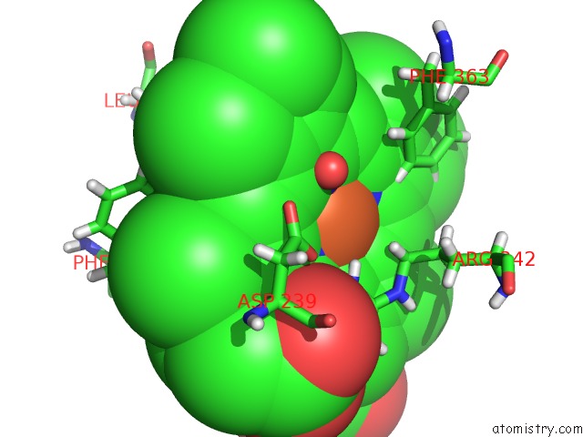
Iron binding site 1 out of 2 in 6i7c
Go back to
Iron binding site 1 out
of 2 in the Dye Type Peroxidase Aa From Streptomyces Lividans: Imidazole Complex
Mono view

Stereo pair view
Mono view
Stereo pair view
A full contact list of Iron with other atoms in the Fe binding
site number 1 of Dye Type Peroxidase Aa From Streptomyces Lividans: Imidazole Complex within 5.0Å range:
|
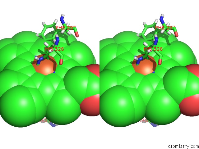
Iron binding site 2 out of 2 in 6i7c
Go back to
Iron binding site 2 out
of 2 in the Dye Type Peroxidase Aa From Streptomyces Lividans: Imidazole Complex

Mono view
Stereo pair view
Mono view
Stereo pair view
A full contact list of Iron with other atoms in the Fe binding
site number 2 of Dye Type Peroxidase Aa From Streptomyces Lividans: Imidazole Complex within 5.0Å range:
|
Reference:
T.Moreno-Chicano,
A.Ebrahim,
D.Axford,
M.V.Appleby,
J.H.Beale,
A.K.Chaplin,
H.M.E.Duyvesteyn,
R.A.Ghiladi,
S.Owada,
D.A.Sherrell,
R.W.Strange,
H.Sugimoto,
K.Tono,
J.A.R.Worrall,
R.L.Owen,
M.A.Hough.
High-Throughput Structures of Protein-Ligand Complexes at Room Temperature Using Serial Femtosecond Crystallography. Iucrj V. 6 1074 2019.
ISSN: ESSN 2052-2525
PubMed: 31709063
DOI: 10.1107/S2052252519011655
Page generated: Tue Aug 6 22:11:06 2024
ISSN: ESSN 2052-2525
PubMed: 31709063
DOI: 10.1107/S2052252519011655
Last articles
Zn in 9MJ5Zn in 9HNW
Zn in 9G0L
Zn in 9FNE
Zn in 9DZN
Zn in 9E0I
Zn in 9D32
Zn in 9DAK
Zn in 8ZXC
Zn in 8ZUF