Iron »
PDB 6i95-6j55 »
6ied »
Iron in PDB 6ied: Crystal Structure of Heme A Synthase From Bacillus Subtilis
Protein crystallography data
The structure of Crystal Structure of Heme A Synthase From Bacillus Subtilis, PDB code: 6ied
was solved by
S.Niwa,
K.Takeda,
M.Kosugi,
E.Tsutsumi,
K.Miki,
with X-Ray Crystallography technique. A brief refinement statistics is given in the table below:
| Resolution Low / High (Å) | 20.00 / 3.00 |
| Space group | H 3 |
| Cell size a, b, c (Å), α, β, γ (°) | 90.528, 90.528, 147.230, 90.00, 90.00, 120.00 |
| R / Rfree (%) | 23.3 / 24.5 |
Other elements in 6ied:
The structure of Crystal Structure of Heme A Synthase From Bacillus Subtilis also contains other interesting chemical elements:
| Copper | (Cu) | 1 atom |
Iron Binding Sites:
The binding sites of Iron atom in the Crystal Structure of Heme A Synthase From Bacillus Subtilis
(pdb code 6ied). This binding sites where shown within
5.0 Angstroms radius around Iron atom.
In total only one binding site of Iron was determined in the Crystal Structure of Heme A Synthase From Bacillus Subtilis, PDB code: 6ied:
In total only one binding site of Iron was determined in the Crystal Structure of Heme A Synthase From Bacillus Subtilis, PDB code: 6ied:
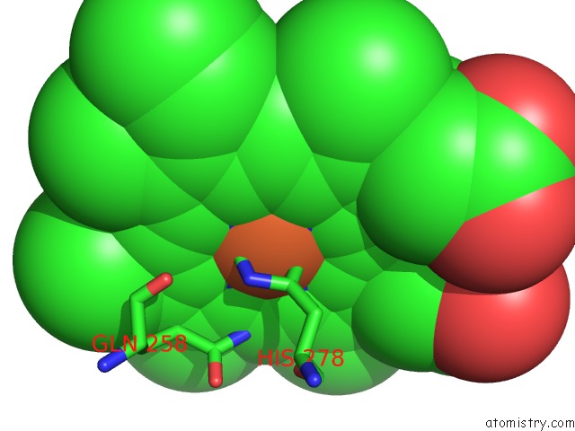
Iron binding site 1 out of 1 in 6ied
Go back to
Iron binding site 1 out
of 1 in the Crystal Structure of Heme A Synthase From Bacillus Subtilis
Mono view

Stereo pair view
Mono view
Stereo pair view
A full contact list of Iron with other atoms in the Fe binding
site number 1 of Crystal Structure of Heme A Synthase From Bacillus Subtilis within 5.0Å range:
|
Reference:
S.Niwa,
K.Takeda,
M.Kosugi,
E.Tsutsumi,
T.Mogi,
K.Miki.
Crystal Structure of Heme A Synthase Frombacillus Subtilis. Proc. Natl. Acad. Sci. V. 115 11953 2018U.S.A..
ISSN: ESSN 1091-6490
PubMed: 30397130
DOI: 10.1073/PNAS.1813346115
Page generated: Wed Aug 6 08:24:16 2025
ISSN: ESSN 1091-6490
PubMed: 30397130
DOI: 10.1073/PNAS.1813346115
Last articles
K in 3T2MK in 3SZB
K in 3SZA
K in 3SYO
K in 3SYQ
K in 3SYP
K in 3SYC
K in 3SYA
K in 3STL
K in 3STZ