Iron »
PDB 6i95-6j55 »
6io9 »
Iron in PDB 6io9: The Structure of Apo-Udgx
Protein crystallography data
The structure of The Structure of Apo-Udgx, PDB code: 6io9
was solved by
W.Xie,
J.Tu,
with X-Ray Crystallography technique. A brief refinement statistics is given in the table below:
| Resolution Low / High (Å) | 36.61 / 1.90 |
| Space group | P 1 21 1 |
| Cell size a, b, c (Å), α, β, γ (°) | 36.572, 50.958, 54.367, 90.00, 104.56, 90.00 |
| R / Rfree (%) | 19 / 22.5 |
Iron Binding Sites:
The binding sites of Iron atom in the The Structure of Apo-Udgx
(pdb code 6io9). This binding sites where shown within
5.0 Angstroms radius around Iron atom.
In total 4 binding sites of Iron where determined in the The Structure of Apo-Udgx, PDB code: 6io9:
Jump to Iron binding site number: 1; 2; 3; 4;
In total 4 binding sites of Iron where determined in the The Structure of Apo-Udgx, PDB code: 6io9:
Jump to Iron binding site number: 1; 2; 3; 4;
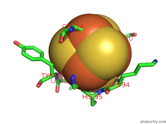
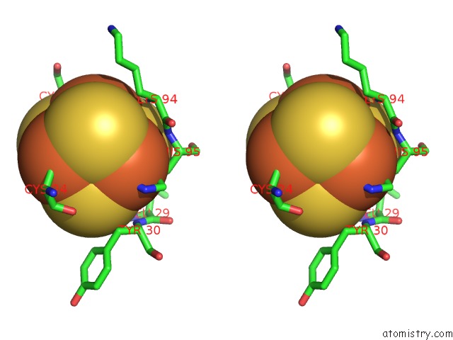
Iron binding site 1 out of 4 in 6io9
Go back to
Iron binding site 1 out
of 4 in the The Structure of Apo-Udgx
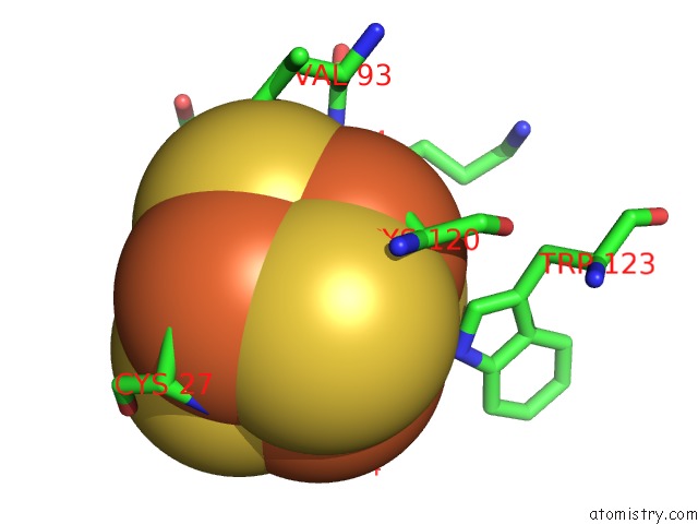
Mono view
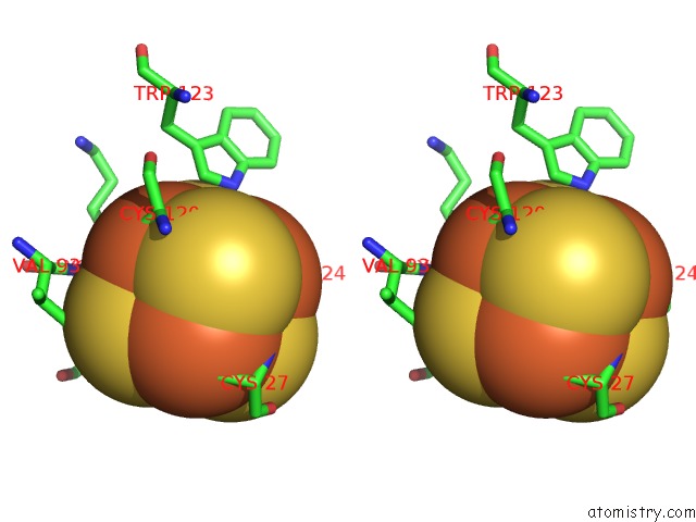
Stereo pair view
Mono view
Stereo pair view
A full contact list of Iron with other atoms in the Fe binding
site number 1 of The Structure of Apo-Udgx within 5.0Å range:
|
Iron binding site 2 out of 4 in 6io9
Go back to
Iron binding site 2 out
of 4 in the The Structure of Apo-Udgx
Mono view
Stereo pair view
Mono view
Stereo pair view
A full contact list of Iron with other atoms in the Fe binding
site number 2 of The Structure of Apo-Udgx within 5.0Å range:
|
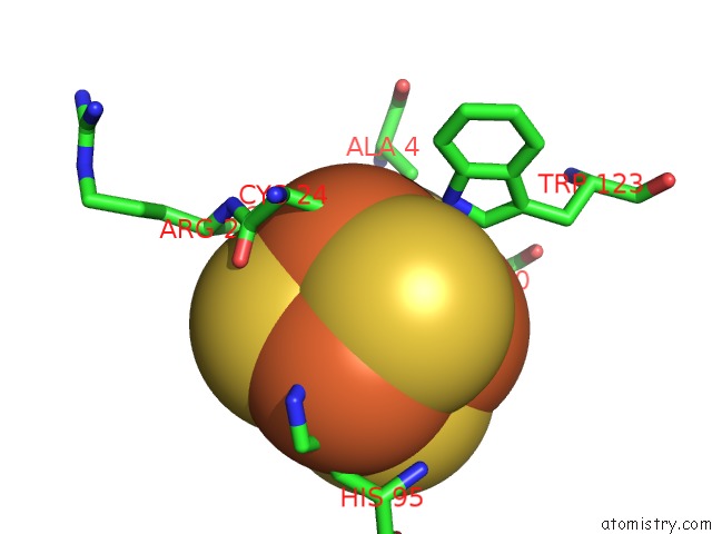
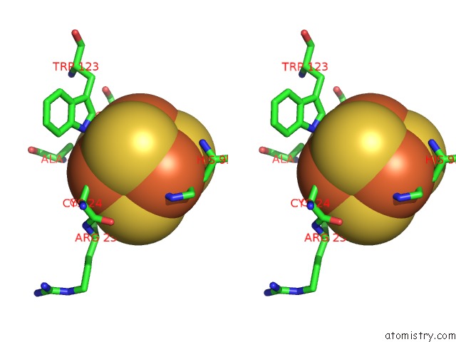
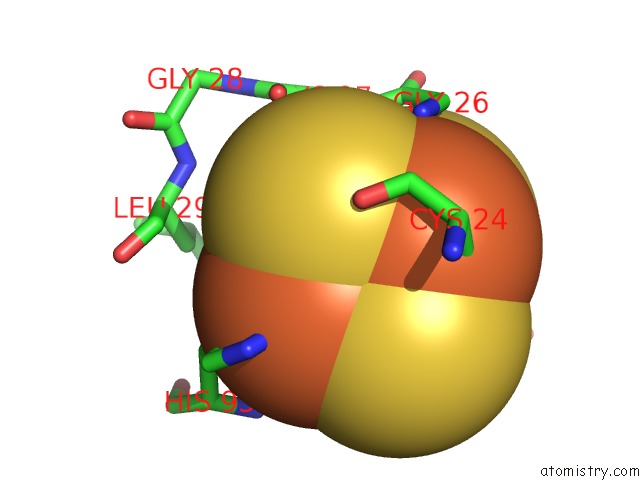
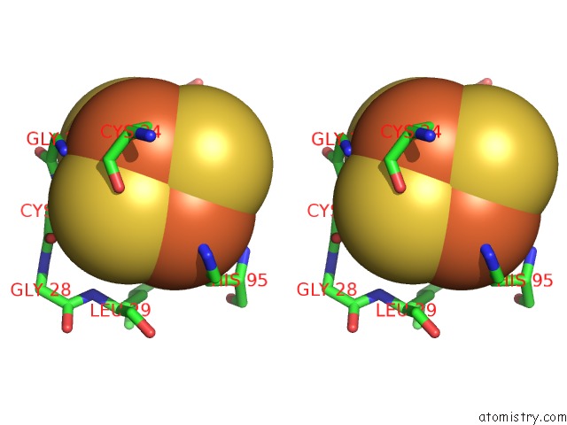
Iron binding site 3 out of 4 in 6io9
Go back to
Iron binding site 3 out
of 4 in the The Structure of Apo-Udgx
Mono view
Stereo pair view
Mono view
Stereo pair view
A full contact list of Iron with other atoms in the Fe binding
site number 3 of The Structure of Apo-Udgx within 5.0Å range:
|
Iron binding site 4 out of 4 in 6io9
Go back to
Iron binding site 4 out
of 4 in the The Structure of Apo-Udgx
Mono view
Stereo pair view
Mono view
Stereo pair view
A full contact list of Iron with other atoms in the Fe binding
site number 4 of The Structure of Apo-Udgx within 5.0Å range:
|
Reference:
J.Tu,
R.Chen,
Y.Yang,
W.Cao,
W.Xie.
Suicide Inactivation of the Uracil Dna Glycosylase Udgx By Covalent Complex Formation. Nat.Chem.Biol. V. 15 615 2019.
ISSN: ESSN 1552-4469
PubMed: 31101915
DOI: 10.1038/S41589-019-0290-X
Page generated: Tue Aug 6 23:03:10 2024
ISSN: ESSN 1552-4469
PubMed: 31101915
DOI: 10.1038/S41589-019-0290-X
Last articles
Fe in 2YXOFe in 2YRS
Fe in 2YXC
Fe in 2YNM
Fe in 2YVJ
Fe in 2YP1
Fe in 2YU2
Fe in 2YU1
Fe in 2YQB
Fe in 2YOO