Iron »
PDB 6l3a-6ll1 »
6l5g »
Iron in PDB 6l5g: Crystal Structure of Yak Lactoperoxidase with Disordered Heme Moiety at 2.50 A Resolution
Protein crystallography data
The structure of Crystal Structure of Yak Lactoperoxidase with Disordered Heme Moiety at 2.50 A Resolution, PDB code: 6l5g
was solved by
P.K.Singh,
C.Rani,
P.Sharma,
S.Sharma,
T.P.Singh,
with X-Ray Crystallography technique. A brief refinement statistics is given in the table below:
| Resolution Low / High (Å) | 64.38 / 2.50 |
| Space group | P 21 21 21 |
| Cell size a, b, c (Å), α, β, γ (°) | 79.820, 84.710, 99.040, 90.00, 90.00, 90.00 |
| R / Rfree (%) | 18.5 / 23.9 |
Other elements in 6l5g:
The structure of Crystal Structure of Yak Lactoperoxidase with Disordered Heme Moiety at 2.50 A Resolution also contains other interesting chemical elements:
| Potassium | (K) | 2 atoms |
| Zinc | (Zn) | 1 atom |
| Calcium | (Ca) | 1 atom |
Iron Binding Sites:
The binding sites of Iron atom in the Crystal Structure of Yak Lactoperoxidase with Disordered Heme Moiety at 2.50 A Resolution
(pdb code 6l5g). This binding sites where shown within
5.0 Angstroms radius around Iron atom.
In total only one binding site of Iron was determined in the Crystal Structure of Yak Lactoperoxidase with Disordered Heme Moiety at 2.50 A Resolution, PDB code: 6l5g:
In total only one binding site of Iron was determined in the Crystal Structure of Yak Lactoperoxidase with Disordered Heme Moiety at 2.50 A Resolution, PDB code: 6l5g:
Iron binding site 1 out of 1 in 6l5g
Go back to
Iron binding site 1 out
of 1 in the Crystal Structure of Yak Lactoperoxidase with Disordered Heme Moiety at 2.50 A Resolution
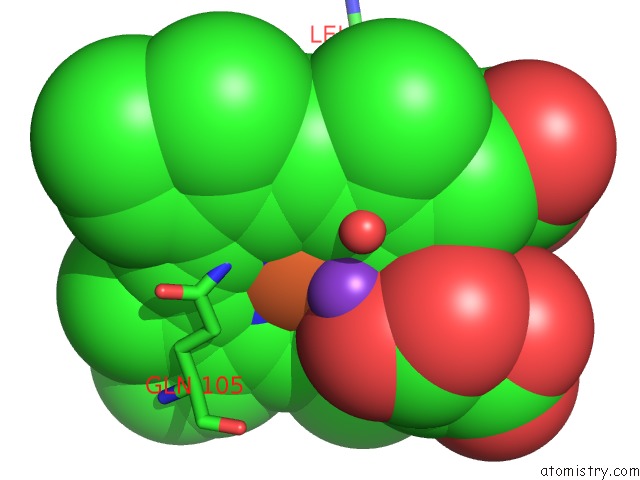
Mono view
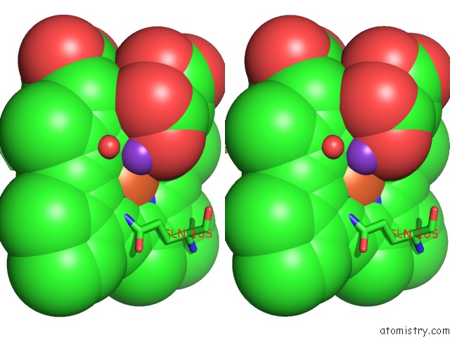
Stereo pair view
Mono view
Stereo pair view
A full contact list of Iron with other atoms in the Fe binding
site number 1 of Crystal Structure of Yak Lactoperoxidase with Disordered Heme Moiety at 2.50 A Resolution within 5.0Å range:
|
Reference:
P.K.Singh,
C.Rani,
P.Sharma,
S.Sharma,
T.P.Singh.
Crystal Structure of Yak Lactoperoxidase with Disordered Heme Moiety at 2.50 A Resolution To Be Published.
Page generated: Wed Aug 6 09:13:25 2025
Last articles
I in 4S3QI in 4TJV
I in 4S22
I in 4S2H
I in 4QX5
I in 4S2G
I in 4S2F
I in 4RX1
I in 4S2D
I in 4RYM